124,99 €
Mehr erfahren.
- Herausgeber: Thieme
- Kategorie: Fachliteratur
- Sprache: Englisch
In the last fifty years, microsystem acupuncture has emerged as a safe and effective therapeutic option for a wide range of conditions, from cardiovascular and neurological disorders to obesity and nicotine dependence. Its worldwide acceptance is largely due to the ability of microsystem acupuncture to provide access for treatment, even in the event of extreme local sensitivity to pain.
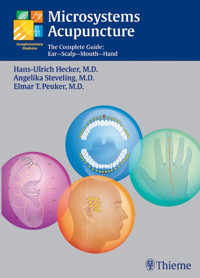
This book is a comprehensive overview of ear, scalp, mouth, and hand acupuncture to help you understand the relationships between these microsystems and the body. Integrating new and established methods, the book covers such topics as Yamamoto New Scalp Acupuncture, Korean hand acupuncture, and laser acupuncture with concise text and more than 350 photographs to allow for instant localization of all zones and points. The range of techniques addressed makes this one-of-a-kind work ideal for busy students and practitioners looking for an overview of some of the most rapidly growing and innovative advances in acupuncture and complementary medicine.
Das E-Book können Sie in Legimi-Apps oder einer beliebigen App lesen, die das folgende Format unterstützen:
Seitenzahl: 310
Veröffentlichungsjahr: 2005
Ähnliche


The Authors
Hans-Ulrich Hecker, M.D.
Medical specialist in general medicine, acupuncture, naturopathy, and homeopathy. Lecturer in Naturopathy and Acupuncture, University of Schleswig-Holstein, Germany. Research Director of Education in Naturopathy and Acupuncture, Academy of Continuing Medical Education of the Regional Medical Association of Schleswig-Holstein.
Certified Medical Quality Manager.
Assessor of the European Foundation of Quality Management (EFQM).
e-mail:praxis@go3docs.de
http:www.go3docs.de
Angelika Steveling, M.D.
Chiropractor, NLP practitioner.
Head of the Department of Traditional Medicine at the Institute for Radiology and Microtherapy, University of Witten-Herdecke, Germany.
Lecturer for Acupuncture Continuing Education, Regional Medical Associations of Schleswig-Holstein and Westphalia-Lippe.
Lecturer of the German Society of Physicians for Acupuncture (DÄGFA)
e-mail: [email protected]
www.akupunktur-ruhr.de
Elmar T. Peuker, M.D.
Medical specialist in general medicine, anatomy, chiropractic, and naturopathy. Lecturer for Acupuncture and Naturopathy Continuing Education, Regional Medical Association of Schleswig-Holstein. Diploma in Health Economy.
Head of the Complementary Medicine Study Group, Department of Anatomy, Wilhelm University of Westphalia, Muenster, Germany.
Lecturer at the British Medical Acupuncture Society (BMAS), UK.
e-mail:[email protected]
www.integrative-medizin.de
For contributors please see page VI.
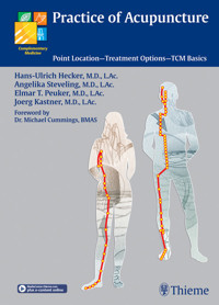
Practice of Acupuncture
The Complete Guide:Ear—Scalp—Mouth—Hand
Hans Ulrich Hecker, M.D., L.Ac.Physician in Private PracticeKiel, Germany
Elmar Peuker, M.D., L.Ac.Clinical AnatomistPhysician in Private PracticeMuenster, Germany
Angelika Steveling, M.D., L.AcPhysician in Private PracticeEssen, Germany
With contributions byMichaela Bijak, John Blank, Timm J. Filler, Hans Garten, Jochen Gleditsch,Bernhard Lichtenauer, Kay Liebchen, Dieter Muehlhoff, Helmut Nissel,Rudolf Rauch, Karen Spiegel, Daniela Stockenhuber, Karsten Strauss,Beate Strittmatter, Max Wiesner-Zechmeister
344 illustrations
ThiemeStuttgart • New York
Library of Congress Cataloging-in-Publication Data
Hecker, Hans-Ulrich.[Lehrbuch und Repetitorium, Ohr-, Schaedel-,Mund-Hand–Akupunktur. English]Microsystems acupuncture: the complete guide: ear–scalp–mouth–hand/Hans Ulrich Hecker, Elmar Peuker, Angelika Steveling; with contributions by Michaela Bijak ... [et al.]; translated by Angela Trowell.
p.; cm.
Authorized and rev. translation of: Lehrbuch und Repetitorium, Ohr-, Schaedel-, Mund–Hand–Akupunktur. 3rd ed. 2002.Includes bibliographical references and index.ISBN 3-13-129111-7 (GTV : alk. paper) - ISBN 1-58890-329-X(TNY : alk. paper)1. Acupuncture points. 2. Acupuncture.I. Peuker, Elmar T. II. Steveling, Angelika. III. Title.[DNLM: 1. Acupuncture Therapy–instrumentation. 2. Acupuncture Therapy–methods. 3. Hand. 4. Head. WB 369 H449L 2006a]RM184.5.H43 2006
615.8’92–dc22 2005023525
This book is an authorized, revised, and expanded translation of the 3rd German edition published and copyrighted 2002 by Hipprokates Verlag, Stuttgart, Germany. Title of the German edition: Lehrbuch und Repetitorium. Ohr-, Schaedel-, Mund-, Hand- Akupunktur. Behandlung ueber das Somatotop
Translator: Angela M. J. Trowell, Granada, Spain
© 2006 Georg Thieme Verlag,Rüdigerstrasse 14, 70469 Stuttgart, Germanyhttp://www.thieme.deThieme New York, 333 Seventh Avenue,New York, NY 10001 USAhttp://www.thieme.com
Typesetting by Martin Wunderlich, KielPrinted in Germany by Appl, Wemding
ISBN 3-13-129111-7 (GTV)ISBN 3-13-129111-7 (TNY) 1 2 3 4 5 6
Important note: Medicine is an ever-changing science undergoing continual development. Research and clinical experience are continually expanding our knowledge, in particular our knowledge of proper treatment and drug therapy. Insofar as this book mentions any dosage or application, readers may rest assured that the authors, editors, and publishers have made every effort to ensure that such references are in accordance with the state of knowledge at the time of production of the book.
Nevertheless, this does not involve, imply, or express any guarantee or responsibility on the part of the publishers in respect to any dosage instructions and forms of applications stated in the book. Every user is requested to examine carefully the manufacturers’ leaflets accompanying each drug and to check, if necessary in consultation with a physician or specialist, whether the dosage schedules mentioned therein or the contraindications stated by the manufacturers differ from the statements made in the present book. Such examination is particularly important with drugs that are either rarely used or have been newly released on the market. Every dosage schedule or every form of application used is entirely at the user’s own risk and responsibility. The authors and publishers request every user to report to the publishers any discrepancies or inaccuracies noticed.
Some of the product names, patents, and registered designs referred to in this book are in fact registered trademarks or proprietary names even though specific reference to this fact is not always made in the text. Therefore, the appearance of a name without designation as proprietary is not to be construed as a representation by the publisher that it is in the public domain.
This book, including all parts thereof, is legally protected by copyright. Any use, exploitation, or commercialization outside the narrow limits set by copyright legislation, without the publisher’s consent, is illegal and liable to prosecution. This applies in particular to photostat reproduction, copying, mimeographing, preparation of microfilms, and electronic data processing and storage.
Preface
For the first time a book has been published which discusses all of the relevant microsystems of acupuncture in practice today. In addition to ear acupuncture, where both Western schools according to Nogier and Bahr as well as Chinese schools are considered, special chapters are given to the following; Chinese Scalp Acupuncture, Yamamoto New Scalp Acupuncture, Mouth Acupuncture, Chinese Hand Acupuncture, Korean Hand Acupuncture, and New Point-Based Pain and Organ Therapy. In addition, the use of laser acupuncture and addiction treatment used with acupuncture is also considered.
The proven team of editors of the Color Atlas of Acupuncture and Practice of Acupuncture has been successful in gaining international recognition as acupuncture specialists through this book. The authors who have contributed to this book have been active in the field of acupuncture training for many years across various disciplines. In many cases, they also teach in universities as lecturers or heads of institutes. Microsystems Acupuncture highlights the most recent views on the diagnoses and therapies used for different somatotopies. The didactical concept, which has been developed by the team of editors and proven in practice, is a guarantee for your learning success.
We would like to thank all of our colleagues who were involved in this book project. We thank Axel Nikolaus for the photographic conversion and Mr Wunderlich for the graphic organization. Last but not least, we give special thanks to our editor, Angelika-Marie Findgott, whose wealth of experience and linguistic authority made the translation and update of this standard textbook possible.
Hans-Ulrich Hecker
Angelika Steveling
Elmar T. Peuker
Contributors
Michaela BijakPhysician in Private PracticeVienna, Austria
John BlankPortland Alternative Health CenterPortland, OR, USA
Timm J. FillerProfessorClinical AnatomistHead of the Clinical Anatomy DivisionUniversity of MuensterMuenster, Germany
Hans GartenAnesthesiologistPhysician in Private PracticeMunich, Germany
Jochen GleditschOtolaryngologist, DentistHonorary President of the German MedicalAcupuncture AssociationBaierbrunn, Germany
Bernhard LichtenauerPhysician in Private PracticeSchwarzau, Austria
Kay LiebchenOrthopedist, RheumatologistPhysician in Private PracticeSchleswig, Germany
Dieter MuehlhoffOncologist, NaturopathPhysician in Private PracticeFelde, Germany
Helmut NisselProfessorDirector of the Kaiserin-Elisabeth-Hospital ViennaVienna, Austria
Rudolf RauchPhysician in Private PracticeVienna, Austria
Karen SpiegelNaturopath, Physician in Private PracticeKiel, Germany
Daniela StockenhuberPhysician in Private PracticePurkersdorf, Austria
Karsten StraussAddiction TherapistInstitute for Addiction MedicineBarkelsby, Germany
Beate StrittmatterNaturopath, Sports medicinePhysician in Private PracticeSpiesen-Elversberg, Germany
Max Wiesner-ZechmeisterPhysician in Private PracticeRied, Austria
Content
1 Basic Principles of Auricular AcupunctureH.-U. Hecker, A. Steveling, E.T. Peuker, B. Strittmatter, T. J. Filler
Introduction
Basic Principles
Zones of Auricular Innervation and Embryological Assignment According to Nogier
Zones of Auricular Innervation According to R.A. Durinjan
More Recent Investigations into Auricular Innervation
Topographic Location of Reflex Zones on the Auricula
Projection of the Skeleton According to Nogier
Topography of Important Projection Zones According to Nogier
Topography of Auricular Acupuncture Points According to Chinese Nomenclature
Topography of Reflex Zones on the Auricula According to R.A. Durinjan
Anatomy of the Rear Side of the Auricula and Projection Zones
The Projection of the Spinal Column in the Region of the Auricula According to Nogier
Significance of Laterality
Rule for the Selection of Auricular Acupuncture Points
Point Searching, Pricking Technique, and Needle Material
2 Topography and Indications of Auricular Acupuncture Points According to RegionsH.-U. Hecker, B. Strittmatter, A. Steveling, E.T. Peuker
Points on the Lobule (1–11) According to Chinese Nomenclature
Points on the Lobule According to Nogier
Points on the Tragus (12–19) and Supratragic Notch (20 and 21) According to Chinese Nomenclature
Points on the Tragus and Supratragic Notch According to Nogier
Points on the Intertragic Notch (Points 22–24) According to Chinese Nomenclature
Points on the Intertragic Notch According to Nogier
Points on the Antitragus (Points 25–36) According to Chinese Nomenclature
Points on the Antitragus According to Nogier
Projection Zones of the Cranial Bones and Sinuses According to Nogier
Points of the Anthelix (Points 37–45) According to Chinese Nomenclature
Projection Zones of the Spinal Column According to Nogier and Bahr
Points on the Superior and Inferior Antihelical Crura (Points 46–54) According to Chinese Nomenclature
Points in the Triangular Fossa (Points 55–61) According to Chinese Nomenclature
Projection Zones of the Lower Extremity According to Nogier
Points in the Region of the Superior and Inferior Anthelical Crura and in the Triangular Fossa According to Nogier
Points on the Scapha (Points 62–71) According to Chinese Nomenclature
Points on the Scapha According to Nogier, Projection of the Upper Extremities
Points on the Scapha According to Nogier
Points on the Helical Rim (Points 72–78) According to Chinese Nomenclature and Nogier
Points on the Ascending Helix Branch (Points 79–83) According to Chinese Nomenclature
Points in the Region of the Ascending Helix Branch (Crus of Helix) According to Nogier, External
Covered Points in the Region of the Ascending Helix Branch (Crus of Helix) According to Nogier
Projection Zones of Internal Organs According to R.A. Durinjan
Projection Zones of Internal Organs According to Chinese Nomenclature
Points around the Helix Root (Points 84–91) According to Chinese Nomenclature
Points in the Superior Concha (Points 92–99) According to Chinese Nomenclature
Points in the Inferior Concha (Points 100–104) According to Chinese Nomenclature
Projection Zones in the Concha (Internal Organs) According to Nogier
Plexus Points and Important Points in the Concha According to Nogier
Points on the Reverse Side of the Auricula (Points 105–108) According to Chinese Nomenclature
Retropoints and Projection of the Spinal Column According to Nogier
Motor Points for Musculature and Joints on the Reverse Side of the Auricula According to Nogier
Motor Points for Thorax and Abdomen on the Left and Right Ear on the Reverse Side of the Auricula According to Nogier
Superordinate Points According to Chinese Nomenclature
Energy and Treatment Lines on the Auricula According to Nogier
Auxiliary Lines in Auricular Acupuncture (B. Strittmatter)
Tutorial: Comparison of the Most Important Auricular Acupuncture Points on the Left and Right Ear
3 Treatment of Major IllnessesH.-U. Hecker, D. Mühlhoff, A. Steveling, E.T. Peuker, K.-H. Junghanns †
Introduction
Internal and Psychosomatic Disorders
(D. Mühlhoff, H.-U. Hecker)
Treatment of Pollinosis
Diseases of the Respiratory Tract
Cardiovascular Diseases
Diseases of the Digestive Organs
Psychosomatic Disorders
Eye Diseases
Dizziness and Tinnitus
Neurological Diseases
Migraine and Cephalgia
Gynecological Disorders (K.-H. Junghanns †)
Urological Diseases
Skin Diseases
4 Diseases of the Locomotor SystemK. Liebchen, H.-U. Hecker
Introduction
Special Clinical Pictures
Cervicogenic Pain Syndrome
Afflictions of the Upper Extremities
Diseases of the Locomotor System: Spinal Column
Diseases of the Hip and Lower Extremities
5 The Medical Treatment of Addiction Using AcupunctureK. Strauss, J. Blank, K. Spiegel
The Use of Acupuncture in the Treatment of Drug-Related Diseases (K. Strauss, J. Blank)
Acupuncture for Nicotine Dependence, Obesity, and Alcohol Dependence (K. Spiegel)
6 Yamamoto New Scalp AcupunctureM. Bijak, D. Stockenhuber, H. Nissel
Presentation of the Method
Implementation
Subdivision of the Somatope
Localization and Indication of the Base Points
Diagnostic Somatopes
YNSA Neck Diagnosis
7 Chinese Scalp AcupunctureH.-U. Hecker, A. Steveling, E.T. Peuker
Introduction
Most Important Projection Zones
Methodology
8 Oral AcupunctureJ. Gleditsch
Introduction
Systematics of Oral Acupuncture
Projection Diagrams of the Retromolar Zones in Quadrants I–II
Projection Diagrams of the Retromolar Zones in Quadrants III–IV
Musculoskeletal System
Pain Management
9 Korean Hand AcupunctureB. Rauch, B. Lichtenauer
Introduction
Technique
Localization Aids for the Palm of the Hand
Hand Acupuncture Points on the Palm of the Hand
Localization Aids for the Dorsal Surface of the Hand
Hand Acupuncture Points on the Dorsal Surface of the Hand
Basic Therapy
Corresponding Therapy
Organ Therapy: Micro-Channels–Ki-Mek Theory
10 Chinese Hand AcupunctureH.-U. Hecker, A. Steveling, E.T. Peuker
Introduction
Technique
Indications and Contraindications
Hand Acupuncture Points on the Dorsal Surface of the Hand
Hand Acupuncture Points on the Palm of the Hand
11 New Point-Based Pain and Organ Therapy (NPPOT)H. Garten
Introduction
Description of Topography
Treatment Technique
Indications
Treatment Areas of Muscular Disturbances and Vertebral Lesions
12 Laser TherapyM. Wiesner-Zechmeister
Use of Low-Level Laser in Fractal Microsystems
The Functional Principle of the Helium–Neon Laser
The Laser Light
Applications of Laser Acupuncture
13 Appendix
Further Reading
Index
Microsystems Acupuncture Today
(J. Gleditsch)
Various Microsystems: Historical Background
Traditional Chinese Medicine (TCM) during the past 50 years has been supplemented and amplified by a new form of acupuncture called microsystem acupuncture.
Microsystem acupuncture is based on particular somatotopic fields comprising specific points of correspondence. Such somatotopic fields were mainly discovered in the West. Microsystems are situated on circumscribed parts of the body, for example, the auricle, the scalp, and the oral cavity. As microsystems resemble cartographies of the organism, they have an allusion to the somatotopic homunculus, as represented at the cerebral hemispheres.
Each of the microsystem points has a clearly defined correlation to, and interrelation with, a particular organ or function. Thus, microsystem acupuncture is a very effective treatment and is established for diagnosis as well.
The first microsystem to be discovered in the early 1950’s was the system of specific points on the auricle. It was the French doctor Nogier who decoded the functional correspondences of the respective auricular points. This punctual cartography resembles a replica of an upside-down embryo. The auricular microsystem is very detailed even though the specific points are densely packed.
Ear acupuncture was continuously refined by Nogier himself as well as by Chinese and Russian schools of acupuncture. Nowadays, auriculotherapy is acknowledged and has gained acceptance worldwide, owing to its therapeutic and diagnostic qualities.
It may be recalled that as early as the close of the 19th century, foot reflexology–probably of Native American Indian origin–had been rediscovered in the U.S.
In the same period, Fliess of Berlin found out that certain digestive, urogenital as well as respiratory disorders responded well when he swabbed specific endonasal zones with a cocaine solution. Obviously, the respective areas of lower and middle nasal conchae were inter-related with specific internal organs and functions. Nasal reflex therapy using specific zones of the nasal mucous membrane was then widely accepted and used by many European practitioners.
Together with auriculotherapy, Yamamoto’s New Scalp Acupuncture (YNSA) has become a very popular form of microsystem acupuncture. In the 1970’s Toshikatsu Yamamoto of Japan discovered various somatotopic zones on the scalp. Specific “basic” zones represent functions of the locomotor system and of the sense organs. In addition, specific “Y”-zones, of 12 points each, represent the respective main channels of TCM. Both basic and Y-zones, as found in the frontal/temporal area, are mirrored once more in the occipital region. Originally, Dr. Yamamoto had discovered striking inter-correlations between the traditional Japanese diagnostic zones of the abdominal wall and specific temporal points. Pain sensitivity and induration of a particular abdominal site is indicative of dysregulation of one of the TCM channels. Therapy applied to a Y-point brings about an immediate dispersal of the corresponding abdominal induration.
Oral acupuncture, also discovered in the 1970’s, is another form of microsystem acupuncture. Intercorrelations of the enoral acupoints are identical with those of the five groups of teeth, as decoded by Voll by means of electro-acupuncture as early as 1965. One particular meridian couple is represented in each one of five dental groups as well as in the adjacent acupoints. In addition to these vestibular points, there are retromolar points, situated beyond the wisdom teeth. These retromolar points are very effective when treating dysfunctions of the locomotor system.
Hand acupuncture has proved to be another effective form of microsystem therapy. During the last decades, Korean Su-Yok (“hand–foot”) acupuncture has become popular in Western countries. In Korean hand acupuncture, the twelve channels as well as reflex points of inner organs and functions and of the skeletal structure are represented by a multitude of points on the palmar and dorsal sides of the hand. A Chinese variant of hand acupuncture provides specific points which are rather related to various indications, with no apparent systematic cartography.
Finally, a somatotopic system situated at the lower leg and foot, discovered by Siener of Germany, has proved effective in therapy.
Characteristics Common to All Microsystems
Common features of microsystem points are: the totality of points comprised in a particular microacu-point system (MAPS) constitutes a functional image of the whole organism in a clearly defined partial area. The respective microsystem points are representative of particular organs and functions and/or of channels of TCM. In this way, microsystem points function as distant points; they always provide treatment, even if a site of pain or dysfunction is not accessible locally. Effects triggered via specific microsystem points are reproducible effects.
After several decades of practice and experience, it has become evident that microsystem therapy works differently to TCM. While the meridian points, owing to the non-stop qi circulation, are constantly available for therapy, in microsystem therapy an “on/off” mechanism is obvious. This results in microsystem points being strictly reactive. They are detectable only in the case of a functional disturbance of the correlated organ. Thus microsystem points show up like “warning signals.”
The activation of microsystem points results in a measurable change of electrical conductivity. This enables bio-electrical point detection. In addition, activated microsystem points are clearly tender to pressure as a rule.
Experience shows that functional disorders are naturally “signaled” simultaneously to analogous points of all microsystems. The degree of point activation, however, may vary from one microsystem to the other. Treatment can be optimized by not sticking to one microsystem only, but by including analogous points of other microsystems as well.
As a rule, if an active point of one microsystem has been treated successfully, this results in analogous points of the other microsystems being deactivated–“deleted”–instantly. Analogous points cease to be detectable.
The “deleting” or “extinguishing” phenomenon indicates a) that a positive therapeutic impulse has been triggered, b) that the choice of points was obviously beneficial, and c) that the patient responds well to acupuncture.
The synonymous terms microsystems, microacu-point systems (MAPS), or somatotopic acupuncture are applicable to each of the following variants:
Systems offering a basically complete organotropic representation of the organism via points or areas of correspondence (e.g. on the auricle, on the soles of the feet).
Systems offering mini-scale representations of the channels depicting every one of the points in a very condensed space (e.g. Korean hand acupuncture).
Systems offering a 12-point representation of the 12 main channels (e.g. YNSA, scalp acupuncture).
Systems offering punctual representation of the respective coupled channel pairs, that is, of the five functional networks (“elements” of TCM), for example, oral acupuncture.
Incomplete micro-point systems specialized in a selection of indications (e.g. nasal reflex zones, Chinese hand acupuncture).
Interestingly, the back shu points, which are representative of the 12 channels, also meet these conditions. In this way, they form a link between TCM and microsystem acupuncture.
The therapeutic effects of acupuncture have been scientifically proven. This applies in particular to pain management achieved by pain research in recent decades. Modulation mechanisms involving endorphin and transmitter activation explain the analgesic effect of both meridian and microsystem acupuncture points. Moreover, spasmolytic, antiphlogistic, sedative, and immunomodulating effects indicate involvement of the autonomic nervous system. According to Bossy, a neuroanatomist at the University of Nîmes, France, it is the reticular formation, where the afferences from the organ in question meet the microsystem point stimuli.
In clinical studies conducted by universities, microsystem points have proved to be superior, particularly on account of their immediate effect, especially in treating locomotor disorders.
Phenomena as seen in microsystem acupuncture may be interpreted in terms of cybernetics and system theory; this applies particularly to mutual networking as well as to the “deleting” phenomenon.
As is known today, the volumes of information, their complexity and networking are increasing in open dissipative systems. An increase in information implies an increase in order. Thus, properties which did not exist previously may emerge, as is the case with nonlinear systems. Fractal geometry, as inaugurated by Mandelbrot, works in the field of nonlinear equations and complex numbers. The recurrence of self-emulating figures is striking when the vast variety of forms is being scaled down progressively. The principle of fractalization (i.e. the similarity principle) has been recognized as the fundamental feature of self-organization in nature. The modern fractal-field model of organism structure opens the way to an understanding of the appearance, structure, and activity of microacupuncture systems.
In living systems, fractalization leads to organisms creating a number of quantum copies of themselves. These replicas seem to provide information exchange between the inner organs and the environment. In terms of cybernetics, therefore, microacupuncture systems are homeostats. The biological significance of these multiple copies is to guarantee greater internal stability and regulation resources.
1 Basic Principles of Auricular Acupuncture
H.-U. Hecker, A. Steveling,E.T. Peuker, B. Strittmatter, T.J. Filler
Introduction
Auricular acupuncture represents a special form of acupuncture and is often used as a complement to body acupuncture.
It is based on a self-contained model of thought. A core idea is the concept of somatopy. This expression is composed of the Greek words soma (= body) and topos (= location) and means the differentiated mapping of the body in one area (here the auricula). Often the term microsystem is used synonymously, although strictly speaking this includes the whole diagnostic and therapeutic concept. Somatopies are familiar from different parts of the central nervous system, for example locomotor somatopy in the Gyrus precentralis or sensory somatopy in the Gyrus postcentralis. By means of somatopy, corresponding constructs are also found for other senses, such as tonotopy for hearing and retinotopy for sight.
As a rule, somatopic assignments are not relative to the size of the mapped region but in accordance with the expression of the respective qualities. Thus, in some cases in the central projection areas there are grotesquely disproportionate representations of the body that are often referred to as homunculus. It is a similar story for the familiar microsystems. The representation of the body on the ear vaguely calls to mind an inverted fetus, the proportions and location of which vary considerably, however, depending on the school of auricular acupuncture (see below).
Unlike body acupuncture, the points on the auricula are only irritated and thus identifiable if there is a disturbance in the respective region of the body of which they are a representative projection. This basic principle also applies to the other microsystem zones.
Therapeutic procedures involving the auricula have been mentioned since antiquity. Thus, Hippocrates is said to have tried to cure impotence by means of bloodletting from the outer ear; Egyptian sailors are said to have tried to improve their sight for navigation by pricking their ear lobes (among other things, the Eye Point in the modern auriculotherapy model is also found in the ear lobe).
Time and time again, cauterization of the auricula was undertaken as a therapy for sciatic pain. We know of corresponding applications by, for example, Persian healers. But in Western Europe there are also references to such therapeutic approaches. Thus, as far back as 1637, Zactus Lusitanus described cauterization of the ear as a therapy for sciatic pain in Portugal; in 1810 Ignaz Colla described cauterization of the rear side of the auricula for the same reason. In 1717, Valsalva treated toothache via the auricula. In the second part (Ling Shu) of the Huang Di Nei Jing, there are observations regarding treatment in the area around the auricula. However, beyond this there are no references to a concept of auricular acupuncture by the standard authors of Chinese medicine.
As early as the 18th century, numerous publications reported the benefits of cauterization of the auricula as a therapy for sciatica. However, it was not until 1950 that the French neurologist Paul Nogier attempted to write a comprehensive description of the therapy of the auricula. He discovered cauterization marks on the anthelix in numerous patients who had been treated by a healer (Mme. Barrin) for sciatic pain. The patients reported astonishing success with this therapy, leading Nogier to further investigate the phenomenon. He also began his own trials with cauterization but then turned to “less barbaric” methods such as pricking with needles or pins, with which he achieved equally good results. He came to the conclusion that disturbances of the body (over and above sciatic pain) could be demonstrated on a regular basis by means of sensitive or painful points on the auricula. He interpreted the representation of the body on the auricula as the image of an inverted fetus–he was thus able to assign the point on the antihelix usually used for sciatic pain therapy the representation zone L4.
In February 1956, at the invitation of the famous acupuncturist Niboyet, Nogier presented his findings at the first congress of the Société méditerranéenne d’Acupuncture in Marseilles, France. At the instance of Gerhard Bachmann (then Chairman of the Deutsche Gesellschaft für Akupunktur [German Society for Acupuncture]), these findings were published in the Deutsche Zeitschrift für Akupunktur (German Acupuncture Journal) in 1957. The findings were not known about in China until the beginning of 1959. Here, although in the past the auricula had been regarded as an important topographical region at which some meridians of body acupuncture meet, independent auricular acupuncture had not previously existed. It was not until the end of 1959 that the expression auricular acupuncture (er zhen) first appeared in Chinese acupuncture literature. Subsequently, “Chinese auricular acupuncture” developed with its own nomenclature and mapping of the ear points which in Europe was employed and interpreted by König and Wancura using a numeric system. Less and less mention is made of the basis of this Chinese auricular acupuncture (the findings of Paul Nogier) in more recent Chinese tutorials.
Nor are there any references in the classic works of Chinese medicine to the microsystems prevalent to a greater or lesser degree in diagnosis and therapy. As a rule, all theories about microsystems are a few years to decades old and have in some cases been implemented retrospectively in one or more of the systems of Chinese medicine.
Taking as a basis the publication of the findings of Paul Nogier in the Deutsche Zeitschrift für Akupunktur, translations or abstracts were also published in Japan, what was then Ceylon (today’s Sri Lanka), and the former USSR.
Today there are several schools of auricular acupuncture; besides the establishment of schools based on Nogier and Bahr on the one hand, and the Chinese school on the other, there has been increasing research activity by Russian scientists based on R.A. Durinjan and F.G. Portnov.
The slight variations in point localizations and approaches in diagnosis and therapy which exist in part between the different schools must not be regarded as rivaling each other, but rather interpreted on the basis of the respective model and the practical approach. Thus, in part, Chinese points represent functional relationships, the Nogier points rather the anatomical correlative. Lastly, a careful investigative technique is crucial for auricular acupuncture in order to identify the individually active points and thus be able to employ them for therapy.
In this book, both the common laws and the differences in the localization of points are described and interpreted, providing the therapist with alternatives which upon closer examination prove to be an enrichment of the range of therapies.
Basic Principles
Anatomy of the Outer Ear (Auricula)
The outer ear together with the auditory canal forms the auricula. Its shape corresponds to the underlying elastic cartilage close to the skin. Only the ear lobe contains no cartilage. Over the dorsomedial area, the skin of the auricula is thin and can be moved relatively easily with regard to the perichondrium. Anterolaterally, the skin is firm and relatively difficult to move.
Both external muscles and own muscles are attached to the auricula. The external muscles permit some people to obtain residual movement of the ear and form part of the mimetic musculature. The ear’s own muscles correspond to the remains of a sphincter system with which the auditory canal can be closed in animals which live in water or underground.
Although the individual internal shape of the ear may vary greatly (and also when the two sides are compared), there are some anatomical landmarks which are relatively constant and can thus serve as reference points for locating the acupuncture points of the ear.
The outer shape of the auricula is formed by the helical rim (helix). The helix originates on the floor of the concha and ascends as the root of helix (crus of helix). It is followed by the body of the helix which descends as the tail of the helix toward the ear lobe. The helix then turns into the ear lobe (auricular lobule). In the upper, rear part of the helix, we usually find a protrusion or widening of the helical rim, the helical tubercle (Darwinian tubercle), which corresponds to the tip of some mammals’ ears. The anthelix runs parallel to the helix. It originates in the upper part of the auricula with two legs, the inferior anthelical crus and the superior anthelical crus. Between the two anthelical crura lies the triangular fossa. The anthelix turns into the antitragus in the lower part of the ear. The border between them is formed by the postantitragal fossa.
Between the helix and the superior anthelical crus plus anthelix lies the scapha.
The tragus is bordered by the intertragic notch to the antitragus and the supratragic notch to the crus of helix. At the bottom of the auricula lies the cavity of concha. The concha is divided by the ascending crus of helix into two parts, the superior concha (cymba) and the inferior concha. The outer auditory canal lies in the inferior concha and is covered from view by the tragus.
The anatomical landmarks of the outside of the auricula partly find their correlate on the rear side. Thus, on the dorsal side of the auricula the helical rim and the eminence of scapha of the sulcus anthelicis can be directly delimited medially. Dorsally both parts of the cavity of concha become the superior and inferior eminence of concha, which are often separated from each other by a sulcus posterior centralis. The equivalents of the crura anthelices (as sulci) and the triangular fossa (as eminentia) on the dorsal side can less frequently be delimited so clearly.
The usually biconcave reverse side of the ear lobe is called the fovea retrobularis.
Zones of Auricular Innervation and Embryological Assignment According to Nogier
Although Nogier did not carry out any anatomical or embryological investigations of his own, several assignments can be found in his records, usually with reference to the research done by Valsalva. According to this, the auricula is innervated by three nerves:
The auricular branch of the vagus nerve
The auriculotemporal nerve of the trigeminal nerve
The great auricular nerve of the cervical plexus.
The auricular branch of the vagus nerve innervates the concha. According to this concept, the “entodermal” organs are projected here.
The great auricular nerve of the cervical plexus supplies the lobule, the outer helical rim up to approximately the Darwinian tubercle, and the back of the ear. These areas correspond to organs in the ectodermal germ layer. The remaining, and by far the largest, part of the ear is innervated by the auriculotemporal nerve of the trigeminal nerve. The mesodermal organs are projected here.
According to Nogier, the different zones are assigned to different functional areas:
Entodermal zoneMetabolism, organs
Mesodermal zoneMotor system
Ectodermal zoneHead and central nervous system
In line with this tripartition, Nogier found one control point for each functional area; these are the Omega Points.
Auriculotemporal nerve(trigeminal nerve)
Auricular branch (vagus nerve)
Great auricular nerve (cervical plexus)
Zones of Auricular Innervation According to Nogier
Zones of Auricular Innervation According to R.A. Durinjan
The description of the auricular zones of innervation and the various somatopic representations according to the Russian school goes back to R.A.Durinjan. The first comprehensive German-language presentation of Russian auriculotherapy came from R. Umlauf and was published in 1988 in the German Journal of Acupuncture (Deutsche Zeitschrift für Akupunktur).
According to Durinjan, the following five nerves participate in the innervation of the auricula:
Fibers of the cervical plexus
The trigeminal nerve,
The intermediate nerve of the facial nerve,
The glossopharyngeal nerve,
The auricular branch of the vagus nerve.
The innervation zones show distinct overlaps of all the areas innervated by the five participating nerves. No auricular zone is therefore exclusively innervated by one single nerve. This might explain why two or more acupuncture points of different functions are projected on identical anatomical sites. Likewise, projections of the same organ are ascribed to different sites of localization. For example, we find projections which correspond to the parenchyma of the organ, next to them projections of the corresponding nervous innervation, and, finally, projections representing the functional state of the organ. Due to the variation in auricular shape, it is conceivable that the overlaps of innervation zones also vary individually. Thus, the frequently described points are really zones rather than points in which the actual ear acupuncture point must be searched for according to individual circumstances. No doubt, this approach goes back to Nogier, who tried to find individual representations of acupuncture points by means of the auriculocardiac reflex (ACR) (cf. p. 30).
Cervical plexus
Glossopharyngeal nerve
Intermediate nerve (facial nerve)
Trigeminal nerve
Vagus nerve
More Recent Investigations into Auricular Innervation
More recent investigations into auricular innervation (Peuker and Filler, 2001) verify a very high density and number of nerve fibers on the external ear compared to other regions of the head. Supply is via four different nerves of both branchiogenic and somatogenic origin:
The great auricular nerve (cervical plexus)
The auriculotemporal nerve (trigeminalnerve)
The auricular branch of the vagus nerve
The occipitalis minor nerve (cervical plexus).
With regard to sensitive innervation, there is a gap between the beginning of the first and third branchiogenic nerve and the third to fifth spinal nerve. There is no overlap between the brianchial arch nerves although there is between the somatogenic nerves and with the brianchial arch nerves.
On the lateral surface, supply via the great auricular nerve (cervical plexus) dominates. The anthelix is mainly supplied solely by the auricular branch of the vagus nerve, in part, by the great auricular nerve, or by both together. The anthelical crura are predominantly innervated by the great auricular nerve. The lobulus and antitragus are usually innervated by the great auricular nerve.
The tragus is mainly supplied jointly by the great auricular nerve and the auriculotemporal nerve. The tail of the helix and scapha are almost always supplied solely by the great auricular nerve; the spina helicis approximately 90% by the auriculotemporal nerve.
The cymba concha is constantly innervated by the auricular branch of the vagus nerve, the cavity of the inferior concha in approximately 50% of cases. In the other 59% of cases, there is dual innervation with the great auricular nerve. There is no overlapping of the zones of innervation of three nerves in any area.
On the dorsal side, the occipitalis minor nerve is involved in innervation, in the upper third often together with the great auricular nerve.
The great auricular nerve and the auricular branch of the vagus nerve are involved in the supply of the middle third, less often the occipitalis minor nerve. The great auricular nerve almost always innervates the lower third, less often the auricular branch of the vagus nerve. There is no area on the dorsal side with threefold innervation either.
Various studies suggest that sensitive innervation of the auricula takes place by means of cranial and cervical nerves (Satomi and Takahashi, 1991). There is still no conclusive explanation for the function of such extensive innervation in man. Possibly, temperature regulation and control of the formation of the ear plays a part here.
Anatomical manuals and atlases examine the nerve supply of the auriculain surprisingly little detail. Most auricular acupuncture manuals are more detailed in this area. However, assumptions with little scientific basis appear to be handed down fromone work to another with regard to the areas of innervation, possibly because in this way hypotheses about postulated modes of action can be supported. An excellent example here is the distinction between entodermal, mesodermal, and ectodermal innervation areas (see above) which cannot be defended for various reasons
Such clearly differentiated innervation definitively does not occur in the human ear.


