35,99 €
Mehr erfahren.
- Herausgeber: John Wiley & Sons
- Kategorie: Fachliteratur
- Serie: At a Glance
- Sprache: Englisch
Highly Commended at the British Medical Association Book Awards 2016
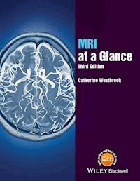
MRI at a Glance encapsulates essential MRI physics knowledge. Illustrated in full colour throughout, its concise text explains complex information, to provide the perfect revision aid. It includes topics ranging from magnetism to safety, K space to pulse sequences, and image contrast to artefacts.
This third edition has been fully updated, with revised diagrams and new pedagogy, including 55 key points, tables, scan tips, equations, and learning points. There is also an expanded glossary and new appendices on optimizing image quality, parameters and trade-offs.
A companion website is also available at www.ataglanceseries.com/mri featuring animations, interactive multiple choice questions, and scan tips to improve your own MRI technique.
MRI at a Glance is ideal for student radiographers and MRI technologists, especially those undertaking the American Registry of Radiation Technologist (ARRT) MRI examination, as well as other health professionals involved in MRI.
Sie lesen das E-Book in den Legimi-Apps auf:
Seitenzahl: 306
Veröffentlichungsjahr: 2015
Ähnliche


This new edition is also available as an e-book.
For more details, please see
www.wiley.com/buy/9781119053552
or scan this QR code:
This edition first published 2016 © 2016 by John Wiley & Sons, Ltd.
Registered office: John Wiley & Sons, Ltd, The Atrium, Southern Gate, Chichester, West Sussex, PO19 8SQ, UK
Editorial offices: 9600 Garsington Road, Oxford, OX4 2DQ, UK 1606 Golden Aspen Drive, Suites 103 and 104, Ames, Iowa 50010, USA
For details of our global editorial offices, for customer services and for information about how to apply for permission to reuse the copyright material in this book please see our website at www.wiley.com/wiley-blackwell
The right of the author to be identified as the author of this work has been asserted in accordance with the UK Copyright, Designs and Patents Act 1988.
All rights reserved. No part of this publication may be reproduced, stored in a retrieval system, or transmitted, in any form or by any means, electronic, mechanical, photocopying, recording or otherwise, except as permitted by the UK Copyright, Designs and Patents Act 1988, without the prior permission of the publisher.
Designations used by companies to distinguish their products are often claimed as trademarks. All brand names and product names used in this book are trade names, service marks, trademarks or registered trademarks of their respective owners. The publisher is not associated with any product or vendor mentioned in this book. It is sold on the understanding that the publisher is not engaged in rendering professional services. If professional advice or other expert assistance is required, the services of a competent professional should be sought.
The contents of this work are intended to further general scientific research, understanding, and discussion only and are not intended and should not be relied upon as recommending or promoting a specific method, diagnosis, or treatment by health science practitioners for any particular patient. The publisher and the author make no representations or warranties with respect to the accuracy or completeness of the contents of this work and specifically disclaim all warranties, including without limitation any implied warranties of fitness for a particular purpose. In view of ongoing research, equipment modifications, changes in governmental regulations, and the constant flow of information relating to the use of medicines, equipment, and devices, the reader is urged to review and evaluate the information provided in the package insert or instructions for each medicine, equipment, or device for, among other things, any changes in the instructions or indication of usage and for added warnings and precautions. Readers should consult with a specialist where appropriate. The fact that an organization or Website is referred to in this work as a citation and/or a potential source of further information does not mean that the author or the publisher endorses the information the organization or Website may provide or recommendations it may make. Further, readers should be aware that Internet Websites listed in this work may have changed or disappeared between when this work was written and when it is read. No warranty may be created or extended by any promotional statements for this work. Neither the publisher nor the author shall be liable for any damages arising herefrom.
Library of Congress Cataloging-in-Publication Data
Westbrook, Catherine. MRI at a glance / Catherine Westbrook. — Third edition. pages cm Includes index. ISBN 978-1-119-05355-2 (pbk.) 1. Magnetic resonance imaging.Outlines, syllabi, etc. 2. Medical physics.Outlines, syllabi, etc. I. Title. RC78.7.N83W4795 2016 616.0-548.dc23
2015022541
A catalogue record for this book is available from the British Library.
Wiley also publishes its books in a variety of electronic formats. Some content that appears in print may not be available in electronic books.
Cover image: © Getty Images/Yuji Sakai
CONTENTS
Preface
Acknowledgement
How to use your textbook
About the companion website
1 Magnetism and electromagnetism
Magnetic susceptibility
Paramagnetism
Diamagnetism
Ferromagnetism
Electromagnetism
2 Atomic structure
Introduction
Motion within the atom
MR active nuclei
3 Alignment
The classical theory
The quantum theory
What do the quantum and classical theories tell us?
4 Precession
Precessional (Larmor) frequency
Precessional phase
5 Resonance and signal generation
Energy absorption
Phase coherence
The MR signal
6 Contrast mechanisms
What is contrast?
Extrinsic contrast parameters
Intrinsic contrast mechanisms
The composition of fat and water
7 Relaxation mechanisms
Relaxation processes
Field inhomogeneities
8 T1 recovery
T1 recovery in fat
T1 recovery in water
Control of T1 recovery
9 T2 decay
T2 decay in fat
T2 decay in water
Control of T2 decay
10 T1 weighting
Typical values
11 T2 weighting
Typical values
12 PD weighting
Typical values
Other types of weighting
13 Conventional spin echo
Mechanisms of CSE
Contrast
Typical values
Uses
14 Fast or turbo spin echo – how it works
Mechanism
Contrast
15 Fast or turbo spin echo – how it is used
Typical values
Uses
16 Inversion recovery
Mechanism
Contrast
Uses
17 Gradient echo – how it works
Mechanism
18 Gradient echo – how it is used
Typical values
Uses
19 The steady state
Echo generation in the steady state
20 Coherent gradient echo
Mechanism
Typical values
Uses
21 Incoherent gradient echo
Mechanism
Typical values
Uses
22 Steady-state free precession
Mechanism
Typical values
Uses
23 Balanced gradient echo
Mechanism
Typical values
Uses
24 Ultrafast sequences
Turbo gradient echo
Echo planar imaging
Typical values
Uses
25 Diffusion and perfusion imaging
Diffusion weighted imaging
Perfusion imaging
26 Functional imaging techniques
Functional MR imaging (fMRI)
Spectroscopy
27 Gradient functions
How gradients work
28 Slice selection
Mechanism
Slice thickness
29 Phase encoding
Mechanism
30 Frequency encoding
Mechanism
31 K space – what is it?
32 K space – how is it filled?
33 K space and image quality
K space – signal and contrast
K space – spatial resolution
34 Data acquisition – frequency
Changing the receive bandwidth
Changing the frequency matrix
35 Data acquisition – phase
36 Data acquisition – scan time
TR
Phase matrix
Number of signal averages (NSA)
Types of acquisition
Reducing scan time
37 K space traversal and pulse sequences
K space traversal in gradient echo
K space traversal in spin echo
K space traversal in single shot
K space traversal in spiral imaging
38 Alternative K space filling techniques
Partial or fractional averaging
Rectangular FOV
Anti-aliasing/over-sampling
Centric imaging
Keyhole imaging
Parallel imaging
39 Signal to noise ratio
Proton density
Coil type and position
TR
TE
Flip angle
Number of signal averages (NSA)
Receive bandwidth
40 Contrast to noise ratio
Administration of contrast
Magnetization transfer contrast
Chemical suppression techniques
Flow techniques
T2 weighting
41 Spatial resolution
Voxel volume and SNR
Voxel volume and resolution
42 Chemical shift artefacts
Appearance
Remedy
Appearance
Remedy
43 Phase mismapping
Appearance
Remedy
44 Aliasing
Frequency aliasing
Phase aliasing
45 Other artefacts
Magnetic susceptibility
Cross-talk
Truncation artefact
Zipper artefact
46 Flow phenomena
Time-of-flight phenomenon
Entry slice phenomenon
Intra-voxel dephasing
47 Time-of-flight MR angiography
Mechanism
Clinical applications
General advantages of TOF MRA
General disadvantages of TOF MRA
48 Phase contrast MR angiography
Mechanism
Clinical uses
49 Contrast-enhanced MR angiography
Mechanism
Administration
Image timing
50 Contrast agents
Gadolinium
Iron oxide
Other contrast agents
51 Magnets
Permanent magnets
Electromagnets
52 Radiofrequency coils
Transmit coils
Receiver coils
RF coil types
53 Gradients and other hardware
Gradients
The pulse control unit
The operator interface
Data storage
54 MR safety – bio-effects
Static magnetic field bio-effects
Time-varying field bio-effects
Site planning
55 MR safety – projectiles
Quenching
Metallic implants and prostheses
Appendix 1(a): The results of optimizing image quality
Appendix 1(b): Parameters and their associated trade-offs
Appendix 2: Artefacts and their remedies
Appendix 3: Main manufacturers’ acronyms
Glossary
Index
Eula
List of Tables
Chapter 1
Table 1.1
Table 1.2
Chapter 2
Table 2.1
Table 2.2
Chapter 3
Table 3.1
Table 3.2
Chapter 4
Table 4.1
Table 4.2
Table 4.3
Chapter 5
Table 5.1
Table 5.2
Chapter 6
Table 6.1
Table 6.2
Chapter 7
Table 7.1
Table 7.2
Table 7.3
Chapter 8
Table 8.1
Table 8.2
Table 8.3
Chapter 9
Table 9.1
Table 9.2
Table 9.3
Chapter 10
Table 10.1
Table 10.2
Chapter 11
Table 11.1
Table 11.2
Chapter 12
Table 12.1
Table 12.2
Chapter 13
Table 13.1
Table 13.2
Chapter 14
Table 14.1
Table 14.2
Table 14.3
Chapter 15
Table 15.1
Table 15.2
Table 15.3
Chapter 16
Table 16.1
Table 16.2
Chapter 17
Table 17.1
Chapter 18
Table 18.1
Table 18.2
Chapter 19
Table 19.1
Table 19.2
Table 19.3
Chapter 20
Table 20.1
Table 20.2
Table 20.3
Chapter 21
Table 21.1
Table 21.2
Table 21.3
Chapter 22
Table 22.1
Table 22.2
Table 22.3
Chapter 23
Table 23.1
Table 23.2
Table 23.3
Chapter 24
Table 24.1
Table 24.2
Table 24.3
Chapter 25
Table 25.1
Table 25.2
Table 25.3
Chapter 26
Table 26.1
Chapter 27
Table 27.1
Table 27.2
Table 27.3
Chapter 28
Table 28.1
Chapter 29
Table 29.1
Chapter 30
Table 30.1
Chapter 41
Table 41.1
Chapter 42
Table 42.1
Table 42.2
Chapter 43
Table 43.1
Chapter 44
Table 44.1
Chapter 45
Table 45.1
Chapter 51
Table 51.1
Chapter 52
Table 52.1
Chapter 53
Table 53.1
Chapter 54
Table 54.1
Table 54.2
Chapter 55
Table 55.1
List of Illustrations
Chapter 1
Figure 1.1
Paramagnetic properties.
Figure 1.2
Diamagnetic properties.
Figure 1.3
Ferromagnetic properties.
Figure 1.4
The right-hand thumb rule.
Figure 1.5
A simple electromagnet.
Chapter 2
Figure 2.1
The atom.
Figure 2.2
The magnetic moment of the hydrogen 1 nucleus.
Chapter 3
Figure 3.1
Alignment: classical theory.
Figure 3.2
Alignment: quantum theory.
Figure 3.3
The net magnetization vector (NMV).
Chapter 4
Figure 4.1
Precession.
Figure 4.2
Precession of the spin-up and
Figure 4.3
The electromagnetic spectrum.
Figure 4.4
Coherent and incoherent phase positions.
Chapter 5
Figure 5.1
Energy transfer during excitation.
Figure 5.2
The flip angle. What flip angle gives maximum transverse magnetization?
Figure 5.3
Generation of the MR signal. Why would you expect it to be alternating?
Chapter 6
Figure 6.1
An axial image of the brain. Note the difference in contrast between CSF, fat, grey and white matter.
Figure 6.2
A basic pulse sequence showing TR and TE intervals.
Chapter 7
Figure 7.1
Relaxation mechanisms.
Chapter 8
Figure 8.1
The T1 recovery curve
Figure 8.2
T1 recovery in fat.
Figure 8.3
T1 recovery in water.
Figure 8.4
T1 recovery of fat and water.
Figure 8.5
T1 contrast generation.
Chapter 9
Figure 9.1
The T2 decay curve.
Figure 9.2
T2 decay in fat.
Figure 9.3
T2 decay in water.
Figure 9.4
T2 decay curves in fat and water.
Chapter 10
Figure 10.1
Axial T1 weighted image of the brain.
Figure 10.2
Coronal T1 weighted image of the knee.
Figure 10.3
Sagittal T1 weighted image of the lumbar spine.
Chapter 11
Figure 11.1
Axial T2 weighted image of the brain.
Figure 11.2
Axial T2 weighted image of the wrist.
Figure 11.3
Sagittal T2 weighted image of the thoracic spine.
Chapter 12
Figure 12.1
Axial proton density weighted image of the brain.
Figure 12.2
Axial proton density weighted image of the knee.
Figure 12.3
Sagittal proton density weighted image of the ankle.
Chapter 13
Figure 13.1
180° RF rephasing.
Figure 13.2
Single-echo spin echo sequence.
Figure 13.3
Dual-echo spin echo sequence.
Figure 13.4
Coronal T1 weighted SE image of the brachial plexus.
Figure 13.5
Axial T2 weighted SE image of the brain.
Chapter 14
Figure 14.1
The echo train in TSE.
Figure 14.2
Phase encoding versus signal amplitude.
Figure 14.3
K space filling and phase reordering.
Chapter 15
Figure 15.1
Axial T2 weighted TSE image of the abdomen.
Figure 15.2
Axial T1 weighted TSE image of the male pelvis.
Figure 15.3
The fast recovery or ‘DRIVE’ sequence.
Figure 15.4
Fast recovery or ‘DRIVE’ image of the internal auditory meatus.
Chapter 16
Figure 16.1
The inversion recovery sequence
Figure 16.2
T1 weighting in inversion recovery
Figure 16.3
How the use of short TI suppresses the signal from fat in a STIR sequence.
Figure 16.4
Axial T2 weighted FLAIR image of the brain
Figure 16.5
Coronal STIR of the knee.
Chapter 17
Figure 17.1
Flip angle vs signal amplitude.
Figure 17.2
How gradients alter field strength and frequency.
Figure 17.3
How gradients rephase.
Figure 17.4
A basic gradient echo sequence showing how a bipolar application of the frequency-encoding gradient produces a gradient echo.
Chapter 18
Figure 18.1
T1 weighting in gradient echo.
Figure 18.2
Sagittal T1 weighted gradient echo of the ankle.
Figure 18.3
T2* weighting in gradient echo.
Figure 18.4
T2* weighted gradient echo of the four chambers of the heart.
Chapter 19
Figure 19.1
The steady state.
Figure 19.2
Echo formation part 1.
Figure 19.3
Echo formation part 2.
Chapter 20
Figure 20.1
Coherent gradient echo sequence.
Figure 20.2
Echo generation in coherent gradient echo.
Figure 20.3
Axial coherent gradient echo image of the abdomen.
Figure 20.4
Sagittal coherent gradient echo image of the knee with tissue suppression.
Chapter 21
Figure 21.1
Incoherent gradient echo sequence.
Figure 21.2
Echo generation in incoherent gradient echo.
Figure 21.3
Coronal incoherent gradient echo from a 3D data set.
Figure 21.4
Coronal incoherent gradient echo acquired after gadolinium enhancement.
Chapter 22
Figure 22.1
SSFP sequence.
Figure 22.2
Echo generation in SSFP.
Figure 22.3
Axial SSFP image of the brain.
Figure 22.4
Perfusion imaging showing hyper-perfusion within oedema indicating recurrent tumour.
Chapter 23
Figure 23.1
Balanced gradient echo scheme in the balanced gradient echo sequence.
Figure 23.2
Alternating RF pulses balanced gradient echo.
Figure 23.3
Sagittal oblique balanced gradient echo of the cervical cord showing nerve roots and peripheral nerves.
Figure 23.4
Axial balanced gradient echo of the abdomen.
Chapter 24
Figure 24.1
Conventional versus ramped sampling.
Figure 24.2
GE-EPI sequence.
Figure 24.3
SE-EPI sequence.
Figure 24.4
Axial SE-EPI of the abdomen.
Chapter 25
Figure 25.1
Free and restricted diffusion.
Figure 25.2
Axial DWI of the brain showing a left-sided infarct.
Figure 25.3
DTI of the brain showing white matter tracts.
Figure 25.4
Set of perfusion images of the brain.
Chapter 26
Figure 26.1
BOLD images of the brain. Functional areas in red.
Figure 26.2
MR spectra of the brain.
Figure 26.3
Multivoxel MRS technique.
Chapter 27
Figure 27.1
A gradient coil.
Figure 27.2
Gradients and changing field strength.
Figure 27.3
How gradients change frequency and phase.
Figure 27.4
Gradient axes.
Chapter 28
Figure 28.1
Slice selection.
Figure 28.2
Using X, Y and Z gradients to select slices.
Figure 28.3
Transmit bandwidth, gradient slope and slice thickness
Figure 28.4
Timing of slice selection in a spin-echo pulse sequence.
Chapter 29
Figure 29.1
Phase encoding.
Figure 29.2
Steep and shallow phase encodings.
Figure 29.3
Timing of phase encoding in a spin echo pulse sequence.
Chapter 30
Figure 30.1
Frequency encoding.
Figure 30.2
Timing of frequency encoding in a spin echo pulse sequence.
Chapter 31
Figure 31.1
K space lines and numbering.
Figure 31.2
K space and the phase matrix.
Chapter 32
Figure 32.1
K space – the chest of drawers.
Figure 32.2
K space filling in spin echo.
Figure 32.3
Data points in K space.
Chapter 33
Figure 33.1
Phase gradient amplitude vs signal amplitude.
Figure 33.2
K space and signal and resolution data.
Figure 33.3
Image using central K space data points only.
Figure 33.4
K space and signal and resolution data.
Chapter 34
Figure 34.1
The Nyquist theorem.
Figure 34.2
Sampling time and the TE.
Chapter 35
Figure 35.1
Phase-encoding slope and phase shift.
Figure 35.2
The phase curve.
Figure 35.3
Fast Fourier transform.
Chapter 36
Figure 36.1
Data acquisition methods.
Figure 36.2
Encoding in a volume acquisition.
Figure 36.3
Axial T2 weighted image of the abdomen. The patient was unable to hold their breath for the duration of the selected scan time, and motion artefact has occurred.
Chapter 37
Figure 37.1
K space traversal in gradient echo.
Figure 37.2
Single-shot K space traversal.
Figure 37.3
Spiral K space traversal.
Chapter 38
Figure 38.1
Partial Fourier.
Figure 38.2
Centric K space filling.
Figure 38.3
Keyhole imaging.
Figure 38.4
Parallel imaging.
Chapter 39
Figure 39.1
Coil placement versus SNR.
Figure 39.2
TE versus SNR.
Figure 39.3
NSA versus SNR.
Figure 39.4
Receive bandwidth versus SNR.
Chapter 40
Figure 40.1
Sagittal (left) and coronal (right) T1 weighted image after contrast showing an ectopic posterior pituitary.
Figure 40.2
Axial slice of the knee from a 3D acquisition using chemical suppression.
Figure 40.3
Phase contrast MR venogram.
Figure 40.4
Axial T2 weighted image of the liver with chemical suppression. There is a good CNR between the liver lesions and normal liver using this technique, although the overall image quality is poor.
Chapter 41
Figure 41.1
Pixel size versus matrix size. Voxels are larger on the lower diagram, which results in a better SNR but poorer resolution than the upper diagram.
Figure 41.2
FOV versus SNR and resolution.
Figure 41.3
Sagittal image using a 10 mm slice thickness.
Figure 41.4
Sagittal image using a 3 mm slice thickness.
Chapter 42
Figure 42.1
Chemical shift and the receive bandwidth.
Figure 42.2
The periodicity of fat and water.
Figure 42.3
Out-of-phase artefact seen as a black line around the abdominal organs.
Chapter 43
Figure 43.1
The cause of phase mismapping from breathing during the acquisition.
Figure 43.2
An axial image of the abdomen during breathing showing phase mismapping.
Figure 43.3
Respiratory compensation and K space.
Figure 43.4
Pre-saturation to reduce flow artefact.
Chapter 44
Figure 44.1
Aliasing or phase wrap.
Figure 44.2
Coronal image of the chest showing aliasing.
Chapter 45
Figure 45.1
Sagittal GE imaging of the knee with metal screws in place. Magnetic susceptibility artefact is clearly seen.
Figure 45.2
Same patient as in Figure 45.1 using a spin echo sequence. The artefact is reduced because RF rephasing corrects for differences in susceptibility between structures.
Figure 45.3
Cross-talk.
Chapter 46
Figure 46.1
The different types of flow.
Figure 46.2
Time-of-flight flow phenomenon.
Figure 46.3
Co-and countercurrent flow.
Figure 46.4
Intra-voxel dephasing.
Chapter 47
Figure 47.1
Presaturation volume relative to the imaging stack.
Figure 47.2
Flow and the imaging volume.
Figure 47.3
3D TOF MRA of a 4-year-old child showing normal appearances.
Figure 47.4
Axial 3D TOF-MRA of the brain acquired at 3 T (left) and 1.5 T (right). Note the enhanced SNR and CNR of the 3 T image.
Chapter 48
Figure 48.1
Bipolar gradients in phase contrast MRA.
Figure 48.2
Flow-encoding axes in phase contrast MRA.
Figure 48.3
VENC settings.
Figure 48.4
PC venogram of the brain.
Chapter 49
Figure 49.1
Coronal CE MRA of the carotid and vertebral arteries.
Figure 49.2
Coronal CE MRA of the chest.
Figure 49.3
Coronal CE MRA of the abdominal vessels.
Figure 49.4
Coronal CE MRA of the iliac arteries showing an arteriovenous malformation.
Chapter 50
Figure 50.1
Tumbling of water molecules.
Figure 50.2
Axial arthrogram of the hip using gadolinium.
Figure 50.3
Coronal T1 weighted image of a small left acoustic neuroma after administration of gadolinium.
Figure 50.4
Axial T1 weighted image of the liver without (left) and with (right) manganese contrast. The enhanced image shows enhancement of normal A B liver so that the liver pathology is darker.
Chapter 51
Figure 51.1
A permanent magnet.
Figure 51.2
A simple electromagnet.
Figure 51.3
A superconducting system.
Figure 51.4
A high field open system.
Chapter 52
Figure 52.1
Spinal phased array coil.
Figure 52.2
Parallel imaging coils.
Chapter 53
Figure 53.1
The MR system.
Figure 53.2
A three-terminal electromagnet used in gradient coils.
Chapter 54
Figure 54.1
Static field in permanent and superconducting systems.
Figure 54.2
The fringe field.
Figure 54.3
The zoning recommended by the American College of Radiology White Paper on MRI safety. Note that there has to be locked access between Zones II and III.
Chapter 55
Figure 55.1
The pulling power of a pair of scissors in a 1.5 T system.
Figure 55.2
Standard labels associated with MR device testing.
Figure 55.3
Patient with an intracranial vascular clip using spin echo (left) and gradient echo MRI (right). Magnetic susceptibility artefact is clearly seen on the gradient echo image.
Guide
Cover
Contents
Preface
Pages
iv
vii
viii
ix
x
xi
xii
xiii
2
3
4
5
6
7
8
9
10
11
12
13
14
15
16
17
18
19
20
21
22
23
24
25
26
27
28
29
30
31
32
33
34
35
36
37
38
39
40
41
42
43
44
45
46
47
48
49
50
51
52
53
54
55
56
58
59
60
61
62
63
64
65
66
67
68
69
70
71
72
73
74
75
76
77
78
79
80
81
82
83
84
85
86
87
88
89
90
91
92
93
94
95
96
97
98
99
100
101
102
103
104
105
106
107
108
109
110
111
112
113
114
115
116
117
118
119
121
122
123
Preface
MRI at a Glance is one of a series of books that presents complex information in an easily accessible format. This series has become famous for its concise text and clear diagrams, which are laid out with text on one page and diagrams relating to the text on the opposite page. In this way all the information on a particular topic is summarized so that the reader has the essential points at their fingertips.
The third edition has been updated with a new companion website that includes some exciting new features. In the book, some chapters have been streamlined and reorganized and there are some updated images and diagrams. Each topic is presented on two pages for easy reference and large subjects have been broken down into smaller sections. In the book and companion website I have included simple explanations and animations, analogies, bulleted lists, simple tables, key points, equations (but only for those who like them), scan tips, ‘Did You Know’ learning points, some questions and answers and plenty of images to aid the understanding of each topic. There are appendices on tradeoffs, acronyms, abbreviations and artefacts. The glossary has also been expanded.
This book is intended to provide a concise overview of essential facts for revision purposes and for those very new to MRI. For more detailed explanations the reader is directed to MRI in Practice and Handbook of MRI Technique. Indeed, the diagrams and images in this book are taken from these other texts and MRI at a Glance is intended to complement them.
Learning MRI physics can be hard work. I hope that this book helps to demystify it!
Acknowledgements
Once again I thank my friend and colleague John Talbot for his beautiful diagrams and for his support. We make a great team and long may it continue! Thanks again to Philips Medical Systems and GE for supplying the images, and to all my friends and family in Brighton, London, Paris, Witney, Leeds, St Augustine, Atlanta and New York.
CW
How to use your textbook
Features contained within your textbook
Each topic is presented in a double-page spread with clear, easy-to-follow diagrams supported by succlnct explanatory text.
Key Point boxes highlight points to remember.
Your textbook id full of photographs, illustrations and tables.
The website icon indicates that you can find accompanying resources on the book's companion website.
About the companion website
Atomic structure
Figure 2.1 The atom.
Figure 2.2 The magnetic moment of the hydrogen 1 nucleus.
Introduction
Atoms make up all matter in the universe and also therefore in the human body. There are approximately 7 octillion (7 × 1027) atoms in the average 70 kg person. Most of the human body (96%) is made up of just four elements. These are hydrogen, oxygen, carbon and nitrogen. Hydrogen is the most common element in the universe and in humans.
The atom consists of the following particles:
Protons
in the nucleus
are positively charged
Neutrons
in the nucleus
have no charge
Electrons
orbit the nucleus
are negatively charged (
Figure 2.1
).
The following terms are used to characterize an atom:
Atomic number:
number of protons in the nucleus and determines the type of element the atoms make up.
Mass number:
sum of the neutrons and protons in the nucleus.
Atoms of the same element having a different mass number are called isotopes. In a stable atom the number of negatively charged electrons equals the number of positively charged protons. Atoms with a deficit or excess number of electrons are called ions and the process of removing electrons from the atom is called ionization. Only certain types of atoms are available to us in Magnetic Resonance Imaging (MRI). These are atoms whose charged nuclei move or spin. This is because a moving electrical charge produces a magnetic field (see Chapter 1).
Motion within the atom
There are three types of motion of particles in the atom:
Negatively charged electrons spinning on their own axis.
Negatively charged electrons orbiting the nucleus.
Particles within the nucleus spinning on their own axes (
Figure 2.1
).
Each type of motion produces a magnetic field (see Chapter 1). In MRI we are concerned with the motion of particles within the nucleus and the nucleus itself.
MR active nuclei
Protons and neutrons spin about their own axis within the nucleus. The direction of spin is random, so that some particles spin clockwise and others anticlockwise.
When a nucleus has an even mass number, the spins cancel each other out so the nucleus has no net spin.
When a nucleus has an odd mass number, the spins do not cancel each other out and the nucleus spins.
As protons have charge, a nucleus with an odd mass number has a net charge as well as a net spin. Due to the laws of electromagnetic induction (see Chapter 1), a moving unbalanced charge induces a magnetic field around itself. The direction and size of the magnetic field are denoted by a magnetic moment (Figure 2.2). The total magnetic moment of the nucleus is the vector sum of all the magnetic moments of protons in the nucleus. The length of the arrow represents the magnitude of the magnetic moment. The direction of the arrow denotes the direction of alignment of the magnetic moment.
Nuclei with an odd number of protons are said to be


