95,99 €
Mehr erfahren.
- Herausgeber: John Wiley & Sons
- Kategorie: Fachliteratur
- Sprache: Englisch
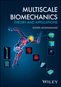
MULTISCALE BIOMECHANICS
Model biomechanical problems at multiple scales with this cutting-edge technology
Multiscale modelling is the set of techniques used to solve physical problems which exist at multiple scales either in space or time. It has been shown to have significant applications in biomechanics, the study of biological systems and their structures, which exist at scales from the macroscopic to the microscopic and beyond, and which produce a myriad of overlapping problems. The next generation of biomechanical researchers therefore has need of the latest multiscale modelling techniques.
Multiscale Biomechanics offers a comprehensive introduction to these techniques and their biomechanical applications. It includes both the theory of multiscale biomechanical modelling and its practice, incorporating some of the latest research and surveying a wide range of multiscale methods. The result is a thorough yet accessible resource for researchers looking to gain an edge in their biomechanical modelling.
Multiscale Biomechanics readers will find:
- Practical biomechanical applications for a variety of multiscale methods
- Detailed discussion of soft and hard tissues, and more
- An introduction to analysis of advanced topics ranging from stenting, drug delivery systems, and artificial intelligence in biomechanics
Multiscale Biomechanics is a useful reference for researchers and scientists in any of the life sciences with an interest in biomechanics, as well as for graduate students in mechanical, biomechanical, biomedical, civil, material, and aerospace engineering.
Sie lesen das E-Book in den Legimi-Apps auf:
Seitenzahl: 671
Veröffentlichungsjahr: 2023
Ähnliche


Multiscale Biomechanics
Theory and Applications
Soheil Mohammadi
School of Civil EngineeringUniversity of TehranTehran, Iran
This edition first published 2023
© 2023 John Wiley & Sons Ltd.
All rights reserved. No part of this publication may be reproduced, stored in a retrieval system, or transmitted, in any form or by any means, electronic, mechanical, photocopying, recording or otherwise, except as permitted by law. Advice on how to obtain permission to reuse material from this title is available at http://www.wiley.com/go/permissions.
The right of Soheil Mohammadi to be identified as the author of this work has been asserted in accordance with law.
Registered Office(s)
John Wiley & Sons, Inc., 111 River Street, Hoboken, NJ 07030, USA
John Wiley & Sons Ltd, The Atrium, Southern Gate, Chichester, West Sussex, PO19 8SQ, UK
For details of our global editorial offices, customer services, and more information about Wiley products visit us at www.wiley.com.
Wiley also publishes its books in a variety of electronic formats and by print-on-demand. Some content that appears in standard print versions of this book may not be available in other formats.
Trademarks
Wiley and the Wiley logo are trademarks or registered trademarks of John Wiley & Sons, Inc. and/or its affiliates in the United States and other countries and may not be used without written permission. All other trademarks are the property of their respective owners. John Wiley & Sons, Inc. is not associated with any product or vendor mentioned in this book.
Limit of Liability/Disclaimer of Warranty
In view of ongoing research, equipment modifications, changes in governmental regulations, and the constant flow of information relating to the use of experimental reagents, equipment, and devices, the reader is urged to review and evaluate the information provided in the package insert or instructions for each chemical, piece of equipment, reagent, or device for, among other things, any changes in the instructions or indication of usage and for added warnings and precautions. While the publisher and authors have used their best efforts in preparing this work, they make no representations or warranties with respect to the accuracy or completeness of the contents of this work and specifically disclaim all warranties, including without limitation any implied warranties of merchantability or fitness for a particular purpose. No warranty may be created or extended by sales representatives, written sales materials or promotional statements for this work. The fact that an organization, website, or product is referred to in this work as a citation and/or potential source of further information does not mean that the publisher and authors endorse the information or services the organization, website, or product may provide or recommendations it may make. This work is sold with the understanding that the publisher is not engaged in rendering professional services. The advice and strategies contained herein may not be suitable for your situation. You should consult with a specialist where appropriate. Further, readers should be aware that websites listed in this work may have changed or disappeared between when this work was written and when it is read. Neither the publisher nor authors shall be liable for any loss of profit or any other commercial damages, including but not limited to special, incidental, consequential, or other damages.
Library of Congress Cataloging-in-Publication Data
Name: Mohammadi, S. (Soheil), author.
Title: Multiscale biomechanics : theory and applications / Soheil Mohammadi
Description: Hoboken, NJ : John Wiley & Sons Ltd., 2023. | Includes bibliographical references and index.
Identifiers: LCCN 2022054751 (print) | LCCN 2022054752 (ebook) | ISBN 9781119033691 (hardback) | ISBN 9781119033738 (pdf) | ISBN 9781119033721 (epub) | ISBN 9781119033714 (ebook)
Subjects: LCSH: Biomechanics.
Classification: LCC QH513 .M642 2023 (print) | LCC QH513 (ebook) | DDC 571.4/3--dc23/eng/20230418
LC record available at https://lccn.loc.gov/2022054751
LC ebook record available at https://lccn.loc.gov/2022054752
Cover image: © SCIEPRO/Getty Images, Courtesy of Soheil Mohammadi
Cover design by Wiley
Set in 9.5/12.5pt STIXTwoText by Integra Software Services Pvt. Ltd, Pondicherry, India
“Love is the unique mysterious solution to the complicated question of Life”
To: Mansoureh, Sogol & Soroush
Contents
Cover
Title Page
Copyright Page
Dedication
Preface
List of Abbreviations
Part I Introduction
1 Introduction
1.1 Introduction to Biomechanics
1.2 Biology and Biomechanics
1.3 Types of Biological Systems
1.3.1 Biosolids
1.3.2 Biofluids
1.3.3 Biomolecules
1.3.4 Synthesized Biosystems
1.4 Biomechanical Hierarchy
1.4.1 Organ Level
1.4.2 Tissue Level
1.4.3 Cellular and Lower Levels
1.4.4 Complex Medical Procedures
1.5 Multiscale/Multiphysics Analysis
1.6 Scope of the Book
Part II Analytical and Numerical Bases
2 Theoretical Bases of Continuum Mechanics
2.1 Introduction
2.2 Solid Mechanics
2.2.1 Elasticity
2.2.2 Plasticity
2.2.3 Damage Mechanics
2.2.4 Fracture Mechanics
2.2.5 Viscoelasticity
2.2.6 Poroelasticity
2.2.7 Large Deformation
2.3 Flow, Convection and Diffusion
2.3.1 Thermodynamics
2.3.2 Fluid Mechanics
2.3.3 Gas Dynamics
2.3.4 Diffusion and Convection
2.4 Fluid–Structure Interaction
2.4.1 Lagrangian and Eulerian Descriptions
2.4.2 Fluid–Solid Interface Boundary Conditions
2.4.3 Governing Equations in the Eulerian Description
2.4.4 Coupled Lagrangian–Eulerian (CLE)
2.4.5 Coupled Lagrangian–Lagrangian (CLL)
2.4.6 Arbitrary Lagrangian–Eulerian (ALE)
3 Numerical Methods
3.1 Introduction
3.2 Finite Difference Method (FDM)
3.2.1 One-Dimensional FDM
3.2.2 Higher Order One-Dimensional FDM
3.2.3 FDM for Solving Partial Differential Equations
3.3 Finite Volume Method (FVM)
3.4 Finite Element Method (FEM)
3.4.1 Basics of FEM Interpolation
3.4.2 FEM Basis Functions/Shape Functions
3.4.3 Properties of the Finite Element Interpolation
3.4.4 Physical and Parametric Coordinate Systems
3.4.5 Main Types of Finite Elements
3.4.6 Governing Equations of the Boundary Value Problem
3.4.7 Numerical Integration
3.5 Extended Finite Element Method (XFEM)
3.5.1 A Review of XFEM Development
3.5.2 Partition of Unity
3.5.3 Enrichments
3.5.4 Signed Distance Function
3.5.5 XFEM Approximation for Cracked Elements
3.5.6 Boundary Value Problem for a Cracked Body
3.5.7 XFEM Discretisation of the Governing Equation
3.5.8 Numerical Integration
3.5.9 Selection of Enrichment Nodes for Crack Propagation
3.5.10 Incompatible Modes of XFEM Enrichments
3.5.11 The Level Set Method for Tracking Moving Boundaries
3.5.12 XFEM Tip Enrichments
3.5.13 XFEM Enrichment Formulation for Large Deformation Problems
3.6 Extended Isogeometric Analysis (XIGA)
3.6.1 Introduction
3.6.2 Isogeometric Analysis
3.6.3 Extended Isogeometric Analysis (XIGA)
3.6.4 XIGA Governing Equations
3.6.5 Numerical Integration
3.7 Meshless Methods
3.7.1 Why Going Meshless
3.7.2 Meshless Approximations
3.7.3 Meshless Solutions for the Boundary Value Problems
3.8 Variable Node Element (VNE)
4 Multiscale Methods
4.1 Introduction
4.2 Homogenization Methods
4.2.1 Introduction
4.2.2 Representative Volume Element (RVE)
4.2.3 Mathematical Homogenization
4.2.4 Computational Homogenization
4.3 Molecular Dynamics (MD)
4.3.1 Introduction
4.3.2 Statistical Mechanics
4.3.3 MD Equations of Motion
4.3.4 Models for Atomic Interactions – MD Potentials
4.3.5 Measures for Determining the State of MD Systems
4.3.6 Stress Computation in MD
4.3.7 Molecular Statics
4.3.8 Sample MD Simulation of a Polymer
4.4 Sequential Multiscale Method
4.4.1 Introduction
4.4.2 Multiscale Modelling of CNT Reinforced Concrete
4.4.3 Molecular Dynamics Simulation of CNTs
4.4.4 Simulation of CNT-Reinforced Calcium Silicate Hydrate
4.4.5 Micromechanical Simulation of CNT-Reinforced Cement
4.4.6 Mesoscale Simulation of CNT-Reinforced Concrete
4.4.7 Macroscale Simulation of CNT-Reinforced Concrete
4.5 Concurrent Multiscale Methods
4.5.1 Introduction
4.5.2 Quasi-Continuum Method (QC)
4.5.3 Bridging Domain Method (BDM)
4.5.4 Bridging Scale Method (BSM)
4.5.5 Disordered Concurrent Multiscale Method (DCMM)
4.5.6 Variable Node Multiscale Method (VNMM)
4.5.7 Enriched Multiscale Method (EMM)
Part III Biomechanical Simulations
5 Biomechanics of Soft Tissues
5.1 Introduction
5.2 Physiology of Soft Tissues
5.2.1 Soft Tissues, Skin
5.2.2 Artery
5.2.3 Heart Leaflet
5.2.4 Brain Tissue
5.3 Hyperelastic Models of Soft Tissues
5.3.1 Introduction
5.3.2 Description of Deformation and Definition of Invariants
5.3.3 Isotropic neo-Hookean Hyperelastic Model
5.3.4 Isotropic Mooney–Rivlin Hyperelastic Model
5.3.5 Hyperelastic Models for Multiscale Simulation of Tendon
5.3.6 Anisotropic Hyperelastic Models for Fibrous Tissues
5.3.7 Polyconvex Undamaged Functions for Fibrous Tissues
5.3.8 Damaged Soft Tissue
5.4 Multiscale Modelling of Undamaged Tendon
5.4.1 Fibril Scale
5.4.2 Fibre Scale
5.4.3 Tissue Scale
5.5 Multiscale Analysis of a Human Aortic Heart Valve
5.5.1 Introduction
5.5.2 Organ Scale Simulation
5.5.3 Simulation in the Tissue Scale
5.5.4 Cell Scale Analysis
5.6 Modelling of Ligament Damage
5.7 Modelling of the Peeling Test: Dissection of the Medial Tissue
5.8 Healing in Damaged Soft Tissue
5.8.1 Introduction
5.8.2 Physical Foundation of Tissue Healing
5.8.3 Solution Procedure
5.8.4 Numerical Analysis
5.9 Hierarchical Multiscale Modelling of a Degraded Arterial Wall
5.9.1 Definition of the Problem
5.9.2 Multiscale Model
5.9.3 Hyperelastic Material Models
5.9.4 Computational Framework of the Hierarchical Multiscale Homogenization
5.9.5 Numerical Results
5.10 Multiscale Modelling of the Brain
5.10.1 Introduction
5.10.2 Biomechanics of the Brain
5.10.3 Multiscale Modelling of the Brain (neo-Hookean Model)
5.10.4 Viscoelastic Modelling of the Brain
6 Biomechanics of Hard Tissues
6.1 Introduction
6.1.1 Hard Tissues
6.1.2 Chemical Composition of Bone
6.1.3 Multiscale Structure of Bone
6.1.4 Bone Remodelling
6.1.5 Contents of the Chapter
6.2 Concepts of Fracture Analysis of Hard Tissues
6.2.1 Numerical Studies of Bone Fracture
6.2.2 Constitutive Response of the Bone
6.2.3 Poroelastic Nature of Bone Tissues
6.2.4 Plasticity and Damage
6.2.5 Hyperelastic Response
6.3 Simulation of the Femur Bone at Multiple Scales
6.3.1 Microscale Simulation of the Trabecular Bone
6.3.2 Two-dimensional XFEM Mesoscale Fracture Simulation of the Cortical Bone
6.3.3 Macroscale Simulation of the Femur
6.4 Healing in Damaged Hard Tissue
6.4.1 Introduction
6.4.2 Physical Foundation of Bone Tissue Healing
6.4.3 Solution Procedure
6.4.4 Numerical Analysis
7 Supplementary Topics
7.1 Introduction
7.2 Shape Memory Alloy (SMA) Stenting of an Artery
7.2.1 Stenting Procedures
7.2.2 SMA Constitutive Equations
7.2.3 Contact Mechanics
7.2.4 Modelling of Stenting
7.2.5 Basics of Modelling
7.3 Multiscale Modelling of the Eye
7.4 Pulsatile Blood Flow in the Aorta
7.4.1 Description of the Problem
7.5 Shape Memory Polymer Drug Delivery System
7.6 Artificial Intelligence in Biomechanics
7.6.1 Artificial Intelligence and Machine Learning
7.6.2 Deep Learning
7.6.3 Physics-Informed Neural Networks (PINNs)
7.6.4 Biomechanical Applications of Artificial Intelligence
References
Index
End User License Agreement
List of Tables
CHAPTER 03
Table 3.1 Basic specifications of the Gauss...
CHAPTER 04
Table 4.1 Constants of the Dreiding potential.
Table 4.2 Constant parameters of the...
Table 4.3 Atomistic scale tensile and...
Table 4.4 Elasticity components Cij(GPa) for the CSH models.
Table 4.5 Nanoscale mechanical properties...
Table 4.6 Reduction of mechanical properties...
Table 4.7 Mechanical properties of the cement paste phases.
Table 4.8 Microscale mechanical properties...
Table 4.9 Mechanical properties of the concrete...
Table 4.10 Average macroscale material properties...
Table 4.11 Comparison of the main specifications...
Table 4.12 Specifications of MS, QC and VNMM models.
Table 4.13 Error of QC and VNMM (with respect to MS).
Table 4.14 Specifications of models of the indentation problem.
CHAPTER 05
Table 5.1 Material properties of the abdominal...
Table 5.2 Material properties of the dog bone sample.
Table 5.3 Material constants of the fibril scale.
Table 5.4 Material constants for the fibre scale.
Table 5.5 Material constants for the tissue scale.
Table 5.6 Hyperelastic material parameters for the organ scale.
Table 5.7 Orientation of collagen fibres in different...
Table 5.8 Comparison of the present and reference...
Table 5.9 Material properties of tissue constituents.
Table 5.10 Comparison of the predicted elongations...
Table 5.11 Comparison of the predicted cell aspect...
Table 5.12 Material properties of the ligament sample.
Table 5.13 Damages in matrix and fibre at the...
Table 5.14 Material properties of the media sample.
Table 5.15 Constants of fibroblast and...
Table 5.16 Constants of collagen/ECM density...
Table 5.17 Constants of the equilibrium equation.
Table 5.18 Initial values of the main variables.
Table 5.19 Material constants for numerical simulations.
Table 5.20 Homogenized properties of the media layer.
Table 5.21 Material constants predicted by the...
Table 5.22 Comparison of the maximum von Mises...
Table 5.23 Material properties of the corpus...
Table 5.24 Neo-Hookean material properties...
Table 5.25 Mooney–Rivlin material...
Table 5.26 Mooney–Rivlin material...
Table 5.27 Comparison of maximum von Mises...
Table 5.28 Comparison of Green–Lagrange strains εGL.
Table 5.29 Elastic and viscoelastic material...
Table 5.30 Viscoelastic material parameters...
Table 5.31 Short-term and long-term stress σz for...
Table 5.32 Long-term stress σz in axon and matrix...
CHAPTER 06
Table 6.1 Elastic constants of the trabecular bone.
Table 6.2 Material properties of the micro...
Table 6.3 Elastic constants of the femur model.
Table 6.4 Constants of the diffusion equations...
Table 6.5 Constants of cartilage matrix density...
Table 6.6 Constants of chondrogenic growth factor...
Table 6.7 Initialization of the main variables.
Table 6.8 Material constants for numerical simulations.
List of Illustrations
CHAPTER 01
Figure 1.1 The brain organ and its nerve cell.
Figure 1.2 Sample biomechanical simulation of...
Figure 1.3 A typical bone with a porous microstructure.
Figure 1.4 An illustration of the process of...
Figure 1.5 Simulations of bones on different scales.
Figure 1.6 Typical bronchiole and biofluid (blood flow) simulations.
Figure 1.7 An illustration of a biomolecular structure.
Figure 1.8 Synthesized systems: a shape memory...
Figure 1.9 Simulation of an aortic heart valve leaflet.
Figure 1.10 Multiple scale structure of soft tissues.
Figure 1.11 Sample modelling of the dissection problem.
Figure 1.12 A variety of cells, from a complex nerve...
Figure 1.13 Modelling of the shape memory stenting procedure.
Figure 1.14 A sequential micro- to macroscale modelling.
Figure 1.15 From macro- to microscale modelling of an eye.
Figure 1.16 Fully coupled macro-micro multiscale homogenization.
Figure 1.17 Computational multiscale...
CHAPTER 02
Figure 2.1 Typical stress–strain responses...
Figure 2.2 Elastoplastic response with perfect...
Figure 2.3 Yield surface and plastic loading.
Figure 2.4 Description of a damaged body...
Figure 2.5 A typical elastoplastic damage behaviour.
Figure 2.6 Equivalent stress and the scalar damage variable.
Figure 2.7 Distribution of stress components...
Figure 2.8 A typical tensile plate with various...
Figure 2.9 Definitions of the Westergaard and Williams...
Figure 2.10 Different modes of crack deformation...
Figure 2.11 Mixed mode crack propagation.
Figure 2.12 Fracture toughness for an...
Figure 2.13 Singular (quarter point)...
Figure 2.14 Definitions of the contour...
Figure 2.15 Different models for approximation...
Figure 2.16 Definition of the crack opening displacement.
Figure 2.17 Stress measures affecting a fatigue problem.
Figure 2.18 Stress-life fatigue description.
Figure 2.19 Strain-life fatigue description.
Figure 2.20 Fatigue crack growth criterion.
Figure 2.21 Descriptions of creep and...
Figure 2.22 Creep and relaxation responses...
Figure 2.23 Creep and relaxation responses...
Figure 2.24 The generalized Maxwell model.
Figure 2.25 The generalized Kelvin model.
Figure 2.26 A typical porous medium.
Figure 2.27 Initial and current configurations...
Figure 2.28 Static fluid pressure.
Figure 2.29 Schematic representation...
Figure 2.30 A typical illustration...
Figure 2.31 Different FSI boundary...
Figure 2.32 Lagrangian, Eulerian and ALE Descriptions.
Figure 2.33 Material, spatial and reference ...
CHAPTER 03
Figure 3.1 One-dimensional representation...
Figure 3.2 Imposition of hinge, clamped...
Figure 3.3 A sample structured grid...
Figure 3.4 Cell and vertex centred...
Figure 3.5 Description of the balance...
Figure 3.6 A typical finite element...
Figure 3.7 One-dimensional linear element.
Figure 3.8 Shape functions of the...
Figure 3.9 Mapping of quadrilateral...
Figure 3.10 Triangular finite elements...
Figure 3.11 Serendipity and Lagrangian...
Figure 3.12 3D brick element in the...
Figure 3.13 A body subjected to body...
Figure 3.14 Positions of quadrature...
Figure 3.15 Definition of the signed distance function.
Figure 3.16 Global, local and polar coordinates at the crack tip.
Figure 3.17 A cracked body subjected...
Figure 3.18 Three methods for numerical integration of a cracked element.
Figure 3.19 Typical nodal enrichment...
Figure 3.20 Standard, enriched...
Figure 3.21 Blending elements...
Figure 3.22 Definition of the...
Figure 3.23 A crack in an electro-mechanical plane strain problem.
Figure 3.24 An edge dislocation and its glide plane.
Figure 3.25 Configuration of a wedge sliding contact.
Figure 3.26 Mesh and control net for a disk of radius 10.
Figure 3.27 A two-dimensional cracked medium.
Figure 3.29 Transformations for...
Figure 3.28 Procedure for selection...
Figure 3.30 Transformation...
Figure 3.31 Comparison of the finite...
Figure 3.32 A typical meshless domain.
Figure 3.33 MLS subdomain and the...
Figure 3.34 A simple procedure to...
Figure 3.35 Enforcement of the boundary...
Figure 3.36 Definitions of the nodal...
Figure 3.37 A local support domain associated...
Figure 3.38 Potential discontinuity between neighbour local support domains.
Figure 3.39 Definitions of radial distances.
Figure 3.40 Integral approximation of a continuous function.
Figure 3.41 Definition of a meshless domain with boundary conditions.
Figure 3.42 Background cell integration for EFG.
Figure 3.43 EFG integration and MLS support domain procedure.
Figure 3.44 Definition of equilibrium domains in MLPG.
Figure 3.45 MLPG integration procedure.
Figure 3.46 FPM support domain.
Figure 3.47 Meshing a model with variable node elements.
Figure 3.48 The number and positions...
Figure 3.49 Shape functions of two nodes...
CHAPTER 04
Figure 4.1 A porous SMA beam solved by...
Figure 4.2 The concept of a representative...
Figure 4.3 An elastic domain with a periodic microstructure.
Figure 4.4 Typical solutions of RVE...
Figure 4.5 The first order computational homogenization.
Figure 4.6 The second order computational homogenization.
Figure 4.7 Problems with non-constant gradients.
Figure 4.8 Comparison of micro- and...
Figure 4.9 Homogenization with a micro-based...
Figure 4.10 A cracked heterogeneous plate....
Figure 4.11 FE mesh for the homogenization...
Figure 4.12 Mode shapes of RVE...
Figure 4.13 Contours of the von...
Figure 4.14 Illustrations of basic...
Figure 4.15 Experimental and MD...
Figure 4.16 Definition of the phase space...
Figure 4.17 Deterministic and probabilistic...
Figure 4.18 Definition of an NVE ensemble.
Figure 4.19 Nose–Hoover thermostat...
Figure 4.20 A typical MD model.
Figure 4.21 Periodic boundary conditions...
Figure 4.22 Variations of interatomic energy...
Figure 4.23 Definitions of variables for two...
Figure 4.24 Definition of the cut off distance...
Figure 4.25 Typical RDF results for an...
Figure 4.26 Schematic representation of the weighting...
Figure 4.27 Initial configuration...
Figure 4.28 Post-relaxation configuration...
Figure 4.29 Post-deformation state...
Figure 4.30 Variations of the...
Figure 4.31 Schematic presentation...
Figure 4.32 A CNT with zigzag and armchair rollings.
Figure 4.33 Topological vacancy and SW defects in CNTs.
Figure 4.34 Single and multi (double)-walled CNTs.
Figure 4.35 Typical tensile deformation and tearing of a CNT.
Figure 4.36 Tensile stress–strain...
Figure 4.37 Definition of relative positions...
Figure 4.38 A typical compressive...
Figure 4.39 Typical deformation...
Figure 4.40 Compressive stress–strain...
Figure 4.41 Compressive stress–strain...
Figure 4.42 The ratio of buckling stress of...
Figure 4.43 The ratio of secondary buckling...
Figure 4.44 The ratio of the elastic modulus...
Figure 4.45 DWCNTs with multiple defects on outer and inner walls.
Figure 4.46 Compressive stress–strain response...
Figure 4.47 Compressive stress–strain response...
Figure 4.48 The ratio of buckling stress of defected...
Figure 4.49 The ratio of the secondary buckling stress...
Figure 4.50 Typical illustration of the CSH globules in nanoscale.
Figure 4.51 Typical nanoscale model of the...
Figure 4.52 Typical longitudinal tensile...
Figure 4.53 Typical CNT crack bridging...
Figure 4.54 Continuum model of the pull-out test.
Figure 4.55 Morse-based stress–strain...
Figure 4.56 Comparison of the force-displacement...
Figure 4.57 Schematic computational model of the...
Figure 4.58 Sample illustration of the computational...
Figure 4.59 Damage distribution in the CNT-reinforced...
Figure 4.60 An illustration of the mesoscale CNT-reinforced...
Figure 4.61 Mesoscale CNT-reinforced concrete samples...
Figure 4.62 Axial strain contours in the tensile...
Figure 4.63 Axial strain contours in the compressive...
Figure 4.64 Spatial distributions of aggregates...
Figure 4.65 Spatial distribution of damage in...
Figure 4.66 Spatial distribution of axial strain...
Figure 4.67 Stress–strain responses of the...
Figure 4.68 Spatial distribution of damage...
Figure 4.69 Spatial distribution of axial...
Figure 4.70 Stress–strain responses...
Figure 4.71 CNT-reinforced concrete samples...
Figure 4.72 Spatial distribution of the...
Figure 4.73 Stress–strain responses...
Figure 4.74 Spatial distribution of the axial...
Figure 4.75 Stress–strain responses...
Figure 4.76 Typical reduction of the penetration...
Figure 4.77 A typical illustration for the finite...
Figure 4.78 A typical fully atomistic modelling...
Figure 4.79 Multiscale domain partitioning into...
Figure 4.80 QC atomistic/FEM modelling strategy...
Figure 4.81 Cluster QC with constrained atoms...
Figure 4.82 Generation of ghost forces due...
Figure 4.83 Definition of nanoindentation...
Figure 4.84 QC and full atomic models...
Figure 4.85 Displacement in the Z direction...
Figure 4.86 Load-penetration response...
Figure 4.87 Domain partitioning into continuum and...
Figure 4.88 The interface zone composed of handshake...
Figure 4.89 Illustration of the weight function...
Figure 4.90 A schematic model of the atomistic...
Figure 4.91 A schematic model of the bridging...
Figure 4.92 Different atomic structures.
Figure 4.93 Schematic model of the micro...
Figure 4.94 Initial and deformed positions...
Figure 4.95 Definition of neighbour elements β.
Figure 4.96 The MD model of amorphous...
Figure 4.97 Energy variations during...
Figure 4.98 Variations of the volume...
Figure 4.99 MD results of the tensile...
Figure 4.100 Different initial finite...
Figure 4.101 Different initial finite...
Figure 4.102 Definition of a multiscale...
Figure 4.103 Variable node multiscale...
Figure 4.104 VNME procedure: initial...
Figure 4.105 Initial configuration...
Figure 4.106 The QC model. Source: Adapted...
Figure 4.107 Variations of the virial...
Figure 4.108 A typical domain with a defect...
Figure 4.109 Mapping...
Figure 4.110 Nano-indentation modelling...
Figure 4.111 QC (top) and VNMM and...
Figure 4.112 Deformed configurations...
Figure 4.113 Comparison of horizontal...
Figure 4.114 Comparison of vertical...
CHAPTER 05
Figure 5.1 Typical composition...
Figure 5.2 Hierarchical structure...
Figure 5.3 Deformation of a collagen...
Figure 5.4 Various stages of healing...
Figure 5.5 Schematic illustration...
Figure 5.6 Aortic valve leaflets. Source:...
Figure 5.7 A typical brain and its microstructure...
Figure 5.8 Microstructure of a typical neuron.
Figure 5.9 Schematic illustration of...
Figure 5.10 A typical tissue sample in the...
Figure 5.11 Typical tensile stress–strain...
Figure 5.12 A biomechanical model in the initial...
Figure 5.13 Definition of two sets of fibres...
Figure 5.14 The hierarchical multiscale structure...
Figure 5.15 The numerical model of the sample...
Figure 5.16 Stress–stretch response...
Figure 5.17 Geometry and finite element...
Figure 5.18 Distribution of damage in the...
Figure 5.19 The hierarchical structure of a tendon.
Figure 5.20 Modelling of the fibril scale...
Figure 5.21 Stress–strain response...
Figure 5.22 Axial (top) and shear (bottom)...
Figure 5.23 Modelling at the fibre scale...
Figure 5.24 Stress–strain response...
Figure 5.25 Stress–strain response...
Figure 5.26 Axial (left) and shear (right)...
Figure 5.27 Modelling at the tissue...
Figure 5.28 Stress–strain response...
Figure 5.29 The multiscale model, from...
Figure 5.30 Geometry of the aortic valve...
Figure 5.31 Uniaxial radial (top) and...
Figure 5.32 Comparison of the results...
Figure 5.33 Partitioning of the leaflet...
Figure 5.34 Finite element model of the...
Figure 5.35 Finite element model and the...
Figure 5.36 Deformations of the leaflet...
Figure 5.37 Deformed shapes of the leaflet...
Figure 5.38 Contours of principal stress at...
Figure 5.39 Critical points of the leaflet...
Figure 5.40 Geometry of the tissue-scale...
Figure 5.41 Single element model of biaxial...
Figure 5.42 Comparison of the biaxial model...
Figure 5.43 Critical elements in fibrosa...
Figure 5.44 Finite element model for RVE...
Figure 5.45 Contour of the deformation...
Figure 5.46 Contour of the maximum...
Figure 5.47 Points 1, 2 and 3 on the...
Figure 5.48 Cell-scale RVE consisting...
Figure 5.49 Finite element model...
Figure 5.50 Maximum principal stress...
Figure 5.51 Comparison of the predicted...
Figure 5.52 The sample of the ligament...
Figure 5.53 Stress–stretch response...
Figure 5.54 Geometry, boundary conditions...
Figure 5.55 Distribution of damage in the...
Figure 5.56 Distribution of damage in the...
Figure 5.57 Force-displacement response...
Figure 5.58 The effect of the characteristic...
Figure 5.59 The effect of the characteristic...
Figure 5.60 Geometry, boundary conditions...
Figure 5.61 Global response of the medial...
Figure 5.62 Distribution of damage in the matrix...
Figure 5.63 Orientations of inactive, active...
Figure 5.64 Distribution of the von Mises...
Figure 5.65 Stages of healing a wound...
Figure 5.66 Force interactions in an...
Figure 5.67 The state of internal...
Figure 5.68 The control volume used...
Figure 5.69 Initial distributions of...
Figure 5.70 A planar strip of wounded skin.
Figure 5.71 The numerical model...
Figure 5.72 Distribution of the fibroblast...
Figure 5.73 Comparison of the distribution...
Figure 5.74 Distribution of the myofibroblast...
Figure 5.75 Distribution of the collagen density...
Figure 5.76 Concentration of the growth factor...
Figure 5.77 Displacement profile over time...
Figure 5.78 A circular damaged skin...
Figure 5.79 The finite element mesh...
Figure 5.80 Initial density distributions...
Figure 5.81 Deformed shapes of the skin...
Figure 5.82 Distribution of the fibroblast...
Figure 5.83 Distribution of the myofibroblast density...
Figure 5.84 Distribution of the collagen density at 2, 7...
Figure 5.85 Distribution of the growth factor at 2, 7...
Figure 5.86 Displacement of the skin tissue during...
Figure 5.87 Comparison of the area change between...
Figure 5.88 Geometry of the simplified model...
Figure 5.89 Composition of the artery wall...
Figure 5.90 Partitioning of the degraded...
Figure 5.91 Definition of simplified...
Figure 5.92 RVE II composed of a ground...
Figure 5.93 GA procedure for determination...
Figure 5.94 Schematic illustration of the...
Figure 5.95 The uniaxial tensile test on...
Figure 5.96 The von Mises stress distribution...
Figure 5.97 The von Mises stress distribution...
Figure 5.98 Descriptions of RVE I and RVE II for...
Figure 5.99 Tensile force-stretch responses...
Figure 5.100 Maximum deformation...
Figure 5.101 Contours of the von Mises...
Figure 5.102 Contours of the von Mises...
Figure 5.103 Contours of the von Mises...
Figure 5.104 Contours of the von Mises...
Figure 5.105 Distribution of the von Mises...
Figure 5.106 Distribution of the von Mises...
Figure 5.107 Slimming of the degraded zone...
Figure 5.108 Typical structures of...
Figure 5.109 Axon fibre in the composite...
Figure 5.110 Three-dimensional image...
Figure 5.111 Three-dimensional finite...
Figure 5.112 Displacement...
Figure 5.113 Contour of the von...
Figure 5.114 Contour of the von...
Figure 5.115 Finite element mesh...
Figure 5.116 Displacement contours on...
Figure 5.117 Contour of the von Mises...
Figure 5.118 Contour of the von Mises...
Figure 5.119 Displacement...
Figure 5.120 Von Mises strain (total)...
Figure 5.121 Von Mises stress (total)...
Figure 5.122 Von Mises strain (total)...
Figure 5.123 Von Mises stress (total)...
Figure 5.124 Short-term von Mises stress...
Figure 5.125 Long-term von Mises stress...
Figure 5.126 Von Mises strain contour...
Figure 5.127 RVE with 52.7% volume...
Figure 5.128 Comparison of the present...
Figure 5.129 Different RVEs with...
Figure 5.130 Step 1%, 3% and 5%...
Figure 5.131 Contours of long term...
Figure 5.132 Contours of long term...
Figure 5.133 Contours of long term...
Figure 5.134 Relaxation of stress...
Figure 5.135 Various strain rates...
Figure 5.136 The effect of strain...
CHAPTER 06
Figure 6.1 Different shapes of...
Figure 6.2 Hierarchical structure...
Figure 6.3 Tropocollagen and microfibril.
Figure 6.4 Assembly of collagen...
Figure 6.5 Array of collagen fibrils.
Figure 6.6 Different assembly patterns...
Figure 6.7 Illustrations of lamella and osteon.
Figure 6.8 Cortical and trabecular bone tissues.
Figure 6.9 Samples of macro-level bones.
Figure 6.10 X-ray scans of a...
Figure 6.11 A typical finite element...
Figure 6.12 Voxel-based hexahedral...
Figure 6.13 Sample XFEM crack propagation...
Figure 6.14 Typical macro- and microscale...
Figure 6.15 Modelling of the femur bone...
Figure 6.16 Voxel-converted finite element...
Figure 6.17 Distribution of the von Mises...
Figure 6.18 Tetrahedral finite element microscale...
Figure 6.19 Distribution of the von Mises stress on the...
Figure 6.20 Extreme stress concentration on the microscale...
Figure 6.21 Stress–strain response...
Figure 6.22 A microstructural sample of cortical bone.
Figure 6.23 Microstructural model of heterogeneous...
Figure 6.24 XFEM mesh of the cortical...
Figure 6.25 Von Mises stress contour...
Figure 6.26 Von Mises stress contour...
Figure 6.27 Force-displacement response...
Figure 6.28 Force-crack opening response...
Figure 6.29 Geometry and the finite...
Figure 6.30 Boundary condition and...
Figure 6.31 Distribution of the von...
Figure 6.32 Distribution of the von...
Figure 6.33 Stages of healing of...
Figure 6.34 A strip of fracture...
Figure 6.35 The numerical model...
Figure 6.36 Initial distributions...
Figure 6.37 Distribution of the MSC...
Figure 6.38 Distribution of the chondrocyte...
Figure 6.39 Distribution of the osteoblast...
Figure 6.40 Distribution of the cartilage...
Figure 6.41 Distribution of the bone matrix...
Figure 6.42 Distribution of the chondrogenic...
Figure 6.43 Distribution of the osteogenic...
CHAPTER 07
Figure 7.1 Typical procedures of stent installation.
Figure 7.2 Nitinol SMA response...
Figure 7.3 Typical finite element...
Figure 7.4 Deformed configurations...
Figure 7.5 Typical distribution...
Figure 7.6 Deformed configurations...
Figure 7.7 Zones of phase transformation...
Figure 7.8 Illustrations of the geometry...
Figure 7.9 Illustrations of the geometry...
Figure 7.10 Finite element mesh of the...
Figure 7.11 Multiscale analysis of the...
Figure 7.12 Geometric model of the...
Figure 7.13 Contour of the velocity...
Figure 7.14 Distribution of normal...
Figure 7.15 The concept of an SMP...
Figure 7.16 Production stages...
Figure 7.17 Drug delivery concept...
Figure 7.18 The thermo-viscoelastic...
Figure 7.19 Variation of the capsule...
Figure 7.20 Variation of the von Mises...
Figure 7.21 A simple illustration of data...
Guide
Cover
Title Page
Copyright Page
Dedication
Table of Contents
Preface
List of Abbreviations
Begin Reading
References
Index
End User License Agreement
Pages
i
ii
iii
iv
v
vi
vii
viii
ix
x
xi
xii
xiii
xiv
xv
xvi
xvii
xviii
xix
xx
1
2
3
4
5
6
7
8
9
10
11
12
13
14
15
16
17
18
19
20
21
22
23
24
25
26
27
28
29
30
31
32
33
34
35
36
37
38
39
40
41
42
43
44
45
46
47
48
49
50
51
52
53
54
55
56
57
58
59
60
61
62
63
64
65
66
67
68
69
70
71
72
73
74
75
76
77
78
79
80
81
82
83
84
85
86
87
88
89
90
91
92
93
94
95
96
97
98
99
100
101
102
103
104
105
106
107
108
109
110
111
112
113
114
115
116
117
118
119
120
121
122
123
124
125
126
127
128
129
130
131
132
133
134
135
136
137
138
139
140
141
142
143
144
145
146
147
148
149
150
151
152
153
154
155
156
157
158
159
160
161
162
163
164
165
166
167
168
169
170
171
172
173
174
175
176
177
178
179
180
181
182
183
184
185
186
187
188
189
190
191
192
193
194
195
196
197
198
199
200
201
202
203
204
205
206
207
208
209
210
211
212
213
214
215
216
217
218
219
220
221
222
223
224
225
226
227
228
229
230
231
232
233
234
235
236
237
238
239
240
241
242
243
244
245
246
247
248
249
250
251
252
253
254
255
256
257
258
259
260
261
262
263
264
265
266
267
268
269
270
271
272
273
274
275
276
277
278
279
280
281
282
283
284
285
286
287
288
289
290
291
292
293
294
295
296
297
298
299
300
301
302
303
304
305
306
307
308
309
310
311
312
313
314
315
316
317
318
319
320
321
322
323
324
325
326
327
328
329
330
331
332
333
334
335
336
337
338
339
340
341
342
343
344
345
346
347
348
349
350
351
352
353
354
355
356
357
358
359
360
361
362
363
364
365
366
367
368
369
370
371
372
373
374
375
376
377
378
379
380
381
382
383
384
385
386
387
388
389
390
391
392
393
394
395
396
397
398
399
400
401
402
403
404
405
406
407
408
409
410
411
412
413
414
415
416
417
418
419
420
421
422
423
424
425
426
427
428
429
430
431
432
433
434
435
436
437
438
439
440
441
442
443
444
445
446
447
448
449
450
451
452
453
454
455
456
457
458
459
460
461
462
463
464
465
466
467
468
469
470
471
472
473
474
475
476
477
478
479
480
481
482
483
484
485
486
487
488
489
490
491
492
493
494
495
496
497
498
499
500
501
502
503
504
505
506
507
508
509
510
511
512
513
514
515
516
517
518
519
520
521
522
523
524
525
526
527
528
529
530
531
532
533
534
535
Preface
Life and space have been the most fascinating scientific concepts that I used to cogitate from my childhood; watching the “Star Trek” series or thinking on the way all living organisms have evolved from non-living atoms and molecules.
As a civil engineer with numerical skills, however, it may seem quite unusual to get involved with biological problems. Nevertheless, the computational mechanics has bridged over the different pillars of science, from buildings to aerospace structures, and from spectacular suspension bridges to intelligent nano additives in biological systems.
It all started about a decade ago, when my former student, Shahrokh Shahi, began his thesis endeavour on multiscale biomechanics. After his graduation, we planned for a book on the subject, although he had to separate from the project to follow his future quests.
Biomechanics is primarily used to study the wide range of mechanical responses of biosystems: from biomolecules scales up to the organ and body levels, and from routine medical procedures to synthesized tissues. The wealth of well-developed mathematical and numerical methods of solving general engineering and physical problems is now available to assist clinical staff and medical industries in assessing the existing procedures and products or to propose new engineered designs and concepts for future research and development.
I have tried to provide the theoretical and computational bases of biomechanics in this textbook and to present my small contribution to encourage young talents to further advance the numerical capabilities for analysis of biomechanical applications.
The book, which can be regarded as an introduction to multiscale biomechanics, is composed of three parts. The preliminary part is meant to provide an introduction and insight into the general concept of biomechanics and the wide variety of biological problems that can be solved numerically through single and multiscale methods.
Part II is dedicated to analytical and numerical bases. In its first set of chapters, the general concepts of continuum mechanics associated with solid materials, fluid flow and diffusion problems are provided. The second set of chapters covers the basics of numerical analysis methods, including the finite element method, the extended finite element method, the isogeometeric analysis, the principles of meshless methods, and the variable node element. The final chapter of Part II discusses the multiscale methods. It begins by examining the homogenization technique. Then the atomistic/molecular dynamics and statistical mechanics are discussed in detail. The sequential multiscale method is thoroughly discussed by a sample case, which spans the extremely wide range of atomistic simulations, nanoscale analysis, microscale computations, mesoscale investigation and macroscale study. The multiscale chapter is concluded by a comprehensive review of concurrent multiscale schemes.
Part III is dedicated to discussions on single and multiscale biomechanical simulations. Its first chapter is devoted to the modelling of soft tissues. It begins with explaining the composition and physiology of soft tissues and their macro and micro hyperelastic constitutive laws. This chapter includes several single and multiscale simulations of soft tissue applications, including damaged tissues, the aortic heart valve, skin damage, arterial wall degradation, wound healing and the viscoelastic response of the brain.
The next chapter of this part is dedicated to hard tissues, and briefly explains the composition of bones and the necessary mechanical models. A number of single and multiscale simulations are presented to provide more insight into the way hard tissue biomechanical studies are performed. The chapter closes with a discussion on the healing processes of hard tissues.
Part III is concluded by a brief complementary chapter on a number of supplementary topics, covering the principles of stenting simulations, multiscale modelling of the eye, the concept of a shape memory polymer drug delivery system and an introduction to artificial intelligence and deep learning in biomechanical applications.
This book is a result of intense research works of the computational community for many years. I have learned from them and tried to pass this package of knowledge to others. I am always obliged to the outstanding works of researchers all over the world and I have done my best to explicitly acknowledge their achievements within the text, figures, tables and formulae, and I sincerely apologize for any unintended failing in appropriately acknowledging them.
I would like to express my acknowledgement to the professional team of John Wiley & Sons, Ltd., for their excellent work that facilitated the whole process of publication of the book: in particular to M. Hammond, E. Willner, A. Hunt, T.P. Koh, S. Lemore, V. Saminathan, D. Shanmugasundram, L. Poplawski and I. Proietti, and to P. Bateson for excellent professional editing of the manuscript.
My sincere thanks to S. Shahi, who jointly began this venture about a decade ago. A number of my colleagues and former students helped me in preparing this book, which I am indebted to acknowledge and sincerely appreciate: Dr. H. Bayesteh reviewed and commented on the homogenization chapter in detail and on time, Dr. O. Alizadeh examined the discussion on concurrent multiscale methods, H. Moslemzadeh prepared the data for a number of sections of the multiscale chapter and the sequential multiscale chapter is credited to Dr. M. Eftekhari. Contributions of M. Vokhshouri and P. Fatemi, and the help of E. Baniasad are acknowledged and the outstanding assistance and contribution of S. Hatefi in different parts are gratefully appreciated.
All numerical simulations of Part III are from the excellent works of my former and present students in the School of Civil Engineering, University of Tehran, beginning with S. Shahi, who developed our basic multiscale solution across multiple levels of organ, tissue and cell, and analysed the heart valve leaflet. Dr. F. Fathi and S. Hatefi then extended the work to damaged soft tissues, and M. Janfada and S. Zolghadr developed the hyperelastic and viscoelastic models of the human brain. In parallel, K. Khaksar solved the complicated coupled system of equations of the healing process in soft tissues, followed by the same approach for hard tissues by M. Zamani. A.R. Torabizadeh eagerly simulated the problems of hard tissues across multiple scales and A. Foyouzat proposed the idea of a nano scale shape memory drug delivery system.
The power for completing the book has been the love, trust and understanding of my beloved family, as they had to comply with all my commitments and to adapt to my long absences. This work is proudly dedicated to noble Iranian students who accomplish academic achievements while challenging for prosperous life and freedom. A living entity may be damaged in harsh environments, but it heals itself, evolves and rises to a flourishing future.
Soheil MohammadiUniversity of TehranSummer–Fall 2022Tehran, Iran
List of Abbreviations
1D
One Dimensional
2D
Two Dimensional
3D
Three Dimensional
AI
Artificial Intelligence
AIS
Abbreviated Injury Scale
ALE
Arbitrary Lagrangian Eulerian
ANN
Artificial Neural Networks
ASME
American Society of Mechanical Engineers
AtC
Atomistic to Continuum Method
AV
Aortic Valve
BCC
Body-Centred Cubic Crystal
BDM
Bridging Domain Method
bFDM
Backward Finite Difference Method
BSM
Bridging Scale Method
CADD
Coupled Atomistics and Discrete Dislocation Mechanics
CAR
Cell Aspect Ratio
CDM
Continuum Damage Mechanics
CEMHYD3D
A Three-Dimensional Cement Hydration and Microstructure Development Modeling Package
CFD
Computational Fluid Dynamics
cFDM
Central Finite Difference Method
CHARMM
Chemistry at Harvard Macromolecular Mechanics (MD Potential)
CLE
Coupled Lagrangian Eulerian
CLL
Coupled Lagrangian-Lagrangian
CNN
Convolutional Neural Networks
CNT
Carbon Nano Tube
CQC
Cluster Quasicontinuum
CSH
Silicate-Cement-Hydrate
CSPM
Corrected SPH Method
CT
Computer Tomography
CTOA
Crack Tip Opening Angle
CTOD
Crack Tip Opening Displacement
DCMM
Disordered Concurrent Multiscale Method
DL
Deep Learning
DNA
Deoxyribonucleic Acid
DOF
Degree of Freedom
DWCNT
Double Wall Carbon Nano Tube
EAM
Embedded Atom Model
ECM
Extracellular Matrix
EDI
Equivalent Domain Integral
EFG
Element Free Galerkin
EMM
Enriched Multiscale Method
EMT
Effective Medium Theory
EPFM
Elastoplastic Fracture Mechanics
FDM
Finite Difference Method
Feat
Finite Element and Atomistic Model
FEM
Finite Element Method
fFDM
Forward Finite Difference Method
FGM
Functionally Graded Material
FPM
Finite Point Method
FS
Finnis–Sinclair
FSI
Fluid-Structure Interaction
FVM
Finite Volume Method
GA
Genetic Algorithm
GAN
Generative Adversarial Networks
HIC
Head Injury Criterion
HPC-Lab
High Performance Computing Laboratory
IC
Interstitial Cell
IGA
Isogeometric Analysis
IKN
Irving–Kirkwood–Noll Stress
IOP
Intraocular Pressure
ITZ
Interface Transition Zone
LAMMPS
Large Scale Atomic/Molecular Massively Parallel Simulator (Software)
LEFM
Linear Elastic Fracture Mechanics
LJ
Lennard–Jones
LSM
Level Set Method
MAAD
Macroscopic Atomistic ab Initio Dynamics
ML
Machine Learning
MD
Molecular Dynamics
MEAM
Modified Embedded Atom Model
MEMS
Micro Electromechanical System
MLPG
Meshless Local Petrov Galerkin
MLS
Moving Least Square
MMP
Matrix Metalloproteinase
MPS
Maximum Principal Strain
MS
Molecular Statics
MSC
Mesenchymal Stem Cell
MSD
Mean Square Displacement Function
MSE
Mean-Squared Error
MWCNT
Multi Wall Carbon Nano Tube
NASGRO
A computer program for analysis of fracture and fatigue crack growth.
NCOP
Non-Collagenous Organic Proteins
NIST
National Institute of Standards and Technology
NiTi
Nickle-Titanium Alloy
NPT
Isothermal-Isobaric Ensemble (Constant Number of Atoms, Pressure and Temperature)
NTG
Normal Tension Glaucoma
NURBS
Non-Uniform Rational B-Spline
NVE
Microcanonical Ensemble (Constant Number of Atoms, Volume and Energy)
NVT
Canonical Ensemble (Constant Number of Atoms, Volume and Temperature)
ONH
Optical Nerve Head
OVITO
Open Visualization Tool
PDE
Partial Differential Equations
PIM
Point Interpolation Method
PINN
Physics-Informed Neural Networks
POAG
Primary Open Angle Glaucoma
PPIM
Polynomial Point Interpolation Method
PTCA
Percutaneous Transluminal Coronary Angioplasty
PU
Partition of Unity
QC
Quasi-continuum
RBF
Radial Basis Function
RDF
Radial Distribution Function
REP
Recovery by Equilibrium on Patch
RKPM
Reproducing Kernel Particle Method
RL
Reinforcement Learning
RNA
Ribonucleic acid
RNN
Recursive (Recurrent) Neural Networks
RPIM
Radial Point Interpolation Method
RPPIM
Radial-Polynomial Point Interpolation Method
RVE
Representative Volume Element
SIF
Stress Intensity Factor
SMA
Shape Memory Alloy
SME
Shape Memory Effect
SMP
Shape Memory Polymer
SPH
Smoothed Particle Hydrodynamics
SPR
Superconvergent Patch Recovery
SW
Stone–Wales Defect
SWCNT
Single Wall Carbon Nano Tube
TBI
Traumatic Brain Injury
TPS
Thin Plate Spline
UV
Ultra Violet
VAF
Velocity Acceleration Function
VNE
Variable Node Element
VNME
Variable Node Multiscale Element
VNMM
Variable Node Multiscale Method
VV
Velocity Verlet
WLS
Weighted Least Square
XFEM
Extended Finite Element Method
XIGA
Extended Isogeometric Analysis
μ-CT
Micro-Computed Tomography
μic
Particle Kinetics Chemical Hydration Model (Software)
Part I Introduction
1 Introduction
1.1 Introduction to Biomechanics
One of the astonishing secrets of life is probably the fact that any living organism is composed of non-living objects: atoms and molecules. Nevertheless, they form biomolecules and eventually constitute the living tissues and organs.
Biology is a comprehensive set of science that is related to living bodies, organs, tissues, cells, molecules and even smaller constituents, and their potential interactions. While biochemistry is part of the biology that is dedicated to what is going on within a biomolecule and their interactions, biomechanics is primarily used to study the wide range of mechanical responses of biosystems; from the biomolecule level up to the organ and body levels.
In this chapter, an introduction is provided to present an insight into the general concept of biomechanics and the wide variety of biological problems that can be solved by conventional or multiscale numerical methods. The numerical approach is widely used in research and development activities for medical-related industries and can be assumed as a reliable tool to assist the medical staff in assessing the existing damages or failures of living tissues or to resemble a specific procedure/operation based on the realistic conditions of a patient and to predict the level of success or potential consequences, before actually performing it.
1.2 Biology and Biomechanics
The word ‘biology’ gradually replaced the phrase of ‘natural history’ well over the centuries to denote the branches of science which deal with living animals or plants (Fung 1993). In fact, it was originally applied to all world-related scientific contributions and not to any individual living object. Nowadays, biology is adopted to designate any science related to the living bodies, organs, tissues, cells, molecules and even smaller constituents, and their potential interactions. It has also spanned to synthetic tissues and includes the technologies of making tailored and engineered materials to replace the existing malfunctioning tissues/organs. The important issue of the drug delivery mechanisms can also be considered within the general concept of biology.
Biomechanics represents an engineering approach to biology, where the general concepts of mechanics are adopted to formulate the biological phenomena and to study them through the wealth of well-developed mathematical and numerical methods of solving general mechanical problems. While some basic mechanical concepts, such as stress or strain, constitutive equations, strength, fatigue and fracture, fluid flow, diffusion, composite action, etc., may not be well defined or used in biology, they are efficiently adopted in biomechanics to define and study the behaviour of living tissues and the existing interactions of different living objects in an organ or in a more complex set of tissues.
Biomechanics spans virtually all aspects of biology, ranging from molecular scales to surgery procedures and from micromodelling of aneurysm disease to the design of large orthopaedic items and haemodialysis machines.
While molecular and cell biology may seem to be only involved with biological and/or chemical phenomena in very small scales, nevertheless, the biomechanics has been adopted to simulate complex organs down to their cell microstructure, such as the brain and its nerve cells, as presented in Figure 1.1, and the heart and its leaflets, the corresponding microscale layers and even an interstitial cell, as depicted in Figure 1.2.
Figure 1.1 The brain organ and its nerve cell.
Figure 1.2 Sample biomechanical simulation of an organ, its tissue layers and the structural molecular/cell level.
It is not always necessary to examine a biomechanical problem down to the cell scale. For instance, in the study of the behaviour of a bone, it is sufficient to take into account its heterogeneous or porous microstructure, as typically presented in Figure 1.3.
Figure 1.3 A typical bone with a porous microstructure.
In surgery-related topics, the whole process of healing of scars or damages of a soft tissue, as typically illustrated in Figure 1.4, can now be fully investigated by biomechanics. These predictions can also be quite useful in clinical orthopaedic or cosmetic surgeries. Similarly, modelling the way a tumour is evolved by the analysis of growth and mass transfer across a membrane/interface to predict how the tumour is contained or expanded by time may help the specialist to obtain a better diagnosis and more effective medical treatment.
Figure 1.4 An illustration of the process of healing in soft tissues.
Biomechanics has been involved in the study of various aspects of the cardiovascular system. For instance, it includes modelling of heart valves, blood flow, hemodynamic disorders and aneurysms, etc. More recently, modelling of the complete procedure of stenting by balloon or through the shape memory effects by detailed macro/microstructural simulation of the blood vessel helps the cardiovascular specialist to decide on a better way for stenting to proceed for any specific patient.
On the other hand, a large number of annual car and industrial accidents and military and terrorist activities have created a growing mass of traumatized and injured people who need orthopaedic replacements in order to get back to a normal life. The corresponding economic impact has justified governments and industries to provide sufficient funding to support the research and development of related biomechanical studies on synthetic biosystems and artificial tissues.
In general, the following steps are involved in any biomechanical computations:
Geometrical macro- and microstructures of the organ/tissue should be determined.
The living organ/tissue performs in a specific environment that should be resembled by biomechanical computations.
The mechanical properties of the constituents must be known. This is probably one of the most challenging parts of a biomechanical analysis due to the size and conditions of a living tissue, which does not readily allow for accurate mechanical tests.
The multiphase and complicated composite nature of the tissue requires a sufficiently accurate model with known constitutive equations.
The results should be verified and calibrated by experiments on the real tissue/organ.
1.3 Types of Biological Systems
Looking at the governing equations and the way the biomechanical systems can be simulated, the following types can be distinguished:
1.3.1 Biosolids
The word biosolid is used to denote solid biological systems. It is noted that biosolid terminology is also used in some public health-related industries to discuss the solid wastes and even toxic chemicals, which are not related to this work.
The biosolid organs and tissues are usually of a composite nature, both in macro- and microscales. While they are solid in nature and are governed by the conventional rules of solid mechanics, they are usually in direct interaction with other types of biomaterials, even with different scales of their biological constituents.
For instance, a soft skin tissue is composed of several distinguishing layers. Some of them have a complex microstructure of collagen fibres within the grounds of a gel-like matrix. The collagen fibre itself is composed of a composite microstructure of fibrils and matrix. The fibres may form a straight or helical shape in different tissues, which constitute the major functionality of the tissue.
As another example, a fracture is a relatively common mode of failure for a bone (Figure 1.5). The bone, as a biosolid, is composed of several layers and a heterogeneous microstructure with different constituents and holes (representing the blood vessels, etc.). Both two- and three-dimensional simulations have been performed to study the fracture resistance of the bone as a single-scale porous solid medium or by advanced multiscale methodology. Such analyses are in practice based on the conventional continuum or fracture mechanics of solids or employ the multiscale analysis of solid materials. Recently, the study of healing of fractured bone has gained a lot of attention and in addition to conventional solid equilibrium equations, requires several coupled equations of the mass transfer and diffusion nature, to be solved simultaneously.
Figure 1.5 Simulations of bones on different scales.
1.3.2 Biofluids
While the earliest written document on blood circulation may be dated back to centuries ago, the breaking work can be attributed to one in the 17th century, where the motion of the heart and blood in animals was discussed for the first time (Harvey 1628), developing the so-called Poiseuille flow and pressure gradient theory in long tubes. It was followed by another major work based on the theory of circulation. Nowadays, massive research studies are being performed on various biofluid applications.
Biofluids (biological flows) cover a wide range of biological applications from organs to cells. Cardiovascular systems, lung and breathing mechanisms, urinary and reproductive systems and even some neurological systems may partially be studied using the biofluid mechanics/dynamics method (Figure 1.6).
Figure 1.6 Typical bronchiole and biofluid (blood flow) simulations.
While the fundamentals of fluid dynamics govern the mechanical responses of biofluids, the well-developed computational fluid dynamics (CFD) can practically and efficiently be used to analyse such systems and to study their interactions with other biological mechanisms. The complexity of the biofluid simulations arise from the fact that in addition to their basic physiological characteristics, their motion and subsequent interactions should also be considered. This is the case when drug delivery conditions are investigated or DNA/RNA and some proteins are sustained in the biofluid.
Moreover, CFD can be combined with conventional solid mechanics and advanced multiscale solutions to study the biomechanical response of different types of diseases such as aneurysms, coronary heart failure and pulmonary problems, leading to better understanding of the corresponding implications, diagnostic and even treatment procedures.
The cardiovascular system is composed of the heart and a blood-carrying network of arteries and veins. The blood flow through the pulmonary and systematic circulation system into the lung arterioles and capillaries allow for the exchange of oxygen and carbon dioxide. The oxygenated blood is then transferred to various organs and cells through systematic circulation. Biofluid mechanics is expected to contribute in the handling of all such complicated mechanisms.
The blood itself is a complex viscous biological fluid, composed of different cells suspended in a plasma, which exchanges oxygen and carbon dioxide within the lungs and other substances within other organs such as kidneys, etc. The plasma contains proteins and other constituents. In addition to the main role of flow mechanics of blood, it performs other roles, such as transfer and diffusion mechanisms of various components that affect the healing process of soft and hard tissues. These effects can be seen in the form of propagation and diffusion processes (Waite and Fine 2007), as will be described in dedicated sections.
1.3.3 Biomolecules
Biomolecules are organic combinations of molecular structures that exist in bio-organisms and contribute to various processes of the bio-object (Figure 1.7). These processes are vital to the existence and performance of living organisms, such as growth factors, differentiation factors, etc.
Figure 1.7 An illustration of a biomolecular structure.
The following biomolecular topics are among the important subjects of research in recent decades:
Proteins are among the primary and essential bases of any biosystem, which affect the life-related functioning of the system.
Lipids, which are mainly fatty acids, are the basic constituent of biological membranes.


