144,99 €
Mehr erfahren.
- Herausgeber: Thieme
- Kategorie: Fachliteratur
- Sprache: Englisch
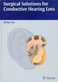
In this book the best surgical solutions for conductive hearing loss due to ossicular fixations are thoroughly described and discussed. The first section covers thympanosclerosis, collating all knowledge on the pathogenesis and pathology of this obscure condition, as well as the surgical methods used in fixation of the ossicular chain, when caused by myringosclerosis and tympanosclerosis. The second section covers postinflammatory, posttraumatic, and postoperative bony fixations of the ossicular chain. In the third section, the voluminous topic of otosclerosis, in particular the evolution of stapes surgery, the various stapedectomy and stapedotomy methods, and the complications of surgery for otosclerosis, are covered in great detail. This section also includes the problems that are encountered in otosclerosis surgery when the ear has been affected with chronic otitis. In the fourth section, congenital ossicular fixations and defects are covered along with some new aspects on the classification and embryology of these problems. For all topics, there is in-depth discussion of the pathogenesis and pathology of the diseases in question. Excellent hand-drawn illustrations provide a level of detail and comprehension not attainable through photography. This book will cover tympanosclerotic, bony, and fibrous fixations, retractions, atelectasis, secretory and adhesive otitis, ventilation problems of the middle ear, cholesteatoma in the tympanic cavity and more!
Das E-Book können Sie in Legimi-Apps oder einer beliebigen App lesen, die das folgende Format unterstützen:
Seitenzahl: 526
Veröffentlichungsjahr: 2000
Ähnliche


Surgical Solutions for Conductive Hearing Loss
Surgical Solutions for Conductive Hearing Loss
Mirko Tos, M.D., Ph.D.
Professor and Chairman
Department of Otorhinolaryngology
Gentofte Hospital
University of Copenhagen
Denmark
Foreword by Ugo Fisch
576 illustrations
ThiemeStuttgart • New York 2000
Library of Congress Cataloging-in-Publication Data
Tos, Mirko.
Surgical solutions for conductive hearing loss / Mirko Tos.
p.; cm.
Includes bibliographical references and index.
ISBN 3131216417 — ISBN 0-86577-910-4
1. Deafness—Surgery. 2. Otosclerosis—Surgery. 3. Ear—Diseases—Surgery. I. Title.
[DNLM: 1. Hearing Loss, Conductive—surgery. 2. Otologic Surgical, Procedures—methods. 3. Otosclerosis—surgery. WV 270 T713s2000]
RF295 .T67 2000
617.8’059—dc21
99-059263
Important Note: Medicine is an ever-changing science undergoing continual development. Research and clinical experience are continually expanding our knowledge, in particular our knowledge of proper treatment and drug therapy. Insofar as this book mentions any dosage or application, readers may rest assured that the authors, editors, and publishers have made every effort to ensure that such references are in accordance with the state of knowledge at the time of production of the book.
Nevertheless, this does not involve, imply, or express any guarantee or responsibility on the part of the publishers in respect of any dosage instructions and forms of application stated in the book. Every user is requested to examine carefully the manufacturer’s leaflets accompanying each drug and to check, if necessary in consultation with a physician or specialist, whether the dosage schedules mentioned there-in or the contraindications stated by the manufacturers differ from the statements made in the present book. Such examination is particularly important with drugs that are either rarely used or have been newly released on the market. Every dosage schedule or every form of application used is entirely at the user’s own risk and responsibility. The authors and publishers request every user to report to the publishers any discrepancies or inaccuracies noticed.
Any reference to or mention of manufacturers or specific brand names should not be interpreted as an endorsement or advertisement for any company or product.
Some of the product names, patents, and registered designs referred to in this book are in fact registered trade-marks or proprietary names even though specific reference to this fact is not always made in the text. Therefore, the appearance of a name without designation as proprietary is not to be construed as a representation by the publisher that it is in the public domain.
This book, including all parts thereof, is legally protected by copyright. Any use, exploitation, or commercialization outside the narrow limits set by copyright legislation, without the publisher’s consent, is illegal and liable to prosecution. This applies in particular to photostat reproduction, copying, mimeographing or duplication of any kind, translating, preparation of microfilms, and electronic data processing and storage.
© 2000 Georg Thieme Verlag, Rüdigerstraße 14, D-70469 Stuttgart, Germany Thieme New York, 333 Seventh Avenue, New York, NY 10001, USA.
Cover design: Martina Berge, Erbach-Ernsbach
Typesetting and Photolitho by BEFORE S.r.l., San Benedetto del Tronto (AP), Italy
Printed in Germany by Grammlich, Pliezhausen
ISBN 3-13-121641-7 (GTV) ISBN 0-86577-910-4 (TNY) eISBN 978-3-1316-2841-1
123456
Foreword
Mirko Tos penetrates with Herculean courage deeper into the intricate forest of the middle ear. His new fascinating journey differs from those of the previous “Manuals” and is made possible by the combination of great surgical experience and a vast knowledge of middle ear pathology. Next to the usual brilliant display of masterly illustrated surgical procedures, the author discusses in great detail the histopathology, pathogenesis, and classification of the tympanosclerotic, posttraumatic, otosclerotic, and congenital ossicular fixations. It is illuminating to realize how this approach gives the reader a better understanding to select the adequate management of difficult surgical problems. It is also refreshing to find in this volume occasional hints as to the preference of the author. All this gives a special touch to this excellent monograph which beautifully reflects all the ingredients of Mirko Tos’ otologic mind.
Every otologist—irrespective of his/her experience—will enjoy this book and look forward to the next one(s). It is reassuring to know that according to classical mythology Hercules successfully accomplished 12 labours!
Zürich, January 2000
Ugo Fisch, M.D. Professor Emeritus ENT Department University Hospital Zürich
Preface
In this book the surgical solutions for conductive hearing loss due to ossicular fixation are described and discussed in detail. It contains four sections. The first deals with tympanosclerosis, collating all knowledge on the pathogenesis and pathology of this obscure condition as all surgical methods used for myringosclerosis and tympanosclerosis fixating the ossicular chain. My research in various aspects of myringosclerosis and tympanosclerosis has a great influence on the monograph. The second deals with postinflammatory, posttraumatic, and postoperative bony fixations of the ossicular chain, explaining its pathogenesis and the surgical methods used in ossicular fixations in the attic and tympanic cavity. Again, my research on ossicular pathology in various middle ear diseases and the dynamics of bone resorption and remodeling has been of great help. The third section summarizes in 13 chapters the enormous subject of otosclerosis, in particular the evolution of stapes surgery, the various stapedectomy and stapedotomy methods, as well as the complications of otosclerosis surgery. The problems encountered in otosclerosis surgery in ears with associated chronic otitis are also included. The fourth section describes in detail, in three chapters, congenital ossicular fixations and defects and presents some new aspects on its classification and embryology.
The illustrations are created in the same way as in my three volumes of Manual of Middle Ear Surgery but involve two artists with different styles, Mrs. Regitze Steinbruch and Mr. Janus Vinther. This came about in the following way: It was my original intention to include all topics of ossicular fixation and all ear canal surgery in Volume 3 of the Manual, and Regitze Steinbruch started with the first eight chapters on fixations. It became obvious, however, that both items could not be included in Volume 3. Therefore, I started to write a separate book on ear canal surgery with Janus Vinther as the artist, who also completed this book. During the last 4 years Janus has been working in my office, allowing an excellent cooperation between the artist and the surgeon. I’ m still indebted to my secretary, Inge Joost, for her great help in typing the manuscript. During the period spent writing these books it became obvious that medicine-related time for a chairman and a chief-secretary is becoming shorter, mainly due to the heavily increasing administrative duties of both. I’ m grateful to Dr. Jonathan Harcourt, ENT surgeon at the Charing Cross Hospital in London, for his great help in language corrections. As with the previous book on ear canal surgery, I appreciated his comments and questions indicating the need for me to express myself more clearly. Kirsti Nielsen and Alis Milman, our hospital librarians, have also been extremely helpful in providing me witch copies of all the literature needed for the book.
Finally, my thanks go to my loving wife, Nives, for supporting me through all four volumes while waiting patiently for my retirement.
In the last paragraph of a preface it has become a tradition to announce and justify a new book with the excuse that several important topics, such as cholesteatoma, adhesive otitis, retractions, atelectasis, and ventilation problems in the middle ear could not be included in this book. The ambition to summarize these topics, being a very important part of my research, in a fifth monograph is still present.
Copenhagen, February 2000
Mirko Tos
Contents
1 Tympanosclerosis
Pathogenesis
Definition
Histopathology
Classification of Tympanosclerosis
Histological Tympanosclerosis
Clinical Tympanosclerosis
Surgical Tympanosclerosis
Pathogenesis of Tympanosclerosis
Stage I: Inflammation and Fibrogenesis
Stage II: Cell Degeneration and Deposition
Stage III: Calcification
Stage IV: Decalcification and Improvement
Myringosclerosis and Grommets
Myringosclerosis and Chronic Otitis
Formation of Middle Ear Tympanosclerosis
2 Surgery of Myringosclerosis
Myringoplasty in Fixation of the Malleus Handle
Inferior Perforation and Anterosuperior Myringosclerosis
Onlay Grafting
Underlay Grafting
Posterior Perforation with Anterior and Inferior Myringosclerosis
Diffuse Massive Myringosclerosis
3 Surgery of Tympanosclerosis
Surgery of Tympanosclerotic Stapes Fixation
Surgical Principles of Stapes Fixation
Tympanosclerosis of the Stapedius Tendon
Moderate and Mild Tympanosclerosis of the Footplate
Moderate Tympanosclerotic Fixation of the Stapes and Footplate
Massive Stapes Tympanosclerosis with an Intact Ossicular Chain
Stapedotomy in Massive Stapes Tympanosclerosis
Stapedectomy with an Intact Ossicular Chain
Stapes Tympanosclerosis with a Disrupted Ossicular Chain
Tympanoplasty Type II with an Intact Stapes
Crurotomy in Massive Stapes Tympanosclerosis
Absent Stapes Crura, Tympanosclerotic Fixated Footplate
Stapedotomy and Stapedectomy with an Absent Incus
Tympanosclerotic Stapes Fixation with an Absent Malleus
Management of Attic Tympanosclerosis
Attic Tympanosclerosis with an Intact Ossicular Chain
Attic and Stapes Tympanosclerosis
4 Bony Ossicular Fixation
Types of Bony Fixations
Malleus Fixation
Bony Fixation of the Incus
Bony Fixation of the Stapes
Incidence of Bony Fixations
Etiology and Pathogenesis
Postinflammatory Pathogenesis
5 Surgery of Bony Fixations
Forcible Mobilization of the Malleus
Atticotomy with Bony Bridge Removal
Atticotomy with Preservation of the Bony Annulus
Atticotomy with Removal of the Bony Annulus
By-pass Technique
Bony Fixation in the Attic and Mobile Stapes
Atticotomy Versus By-pass Technique
Bony Incus Fixation
Bony Fixation of the Stapedial Arch
Resection of the Stapes Crura
Fixed Malleus Associated with Otosclerosis
By-pass Technique
6 Bony Fixation of the Grafts
Bony Fixation to the Posterosuperior Bony Annulus
Bony Fixation to the Facial Nerve Prominence
Bony Fixation to the Promontory and Oval Window Niche
7 Otosclerosis
Definition and Terminology
Classification of Otosclerotic Lesions
Portmann Classification
Gritwood and Causse Classifications
History of Otosclerosis Surgery
The Earliest Period (1878-1900)
Period without Stapes Surgery (1890-1913)
Period with Experimental Fenestration (1913-1938)
The Lempert’s Fenestration Period (1938-1952)
Evolution of Modern Stapes Surgery
Period with Stapes Mobilization (1952-1958)
Stapedoplasty, Stapedectomy, and Stapedotomy Periods (from 1958)
Stapedotomies or Small Fenestra Techniques
Medical Treatment
8 Stapedectomy Techniques
Shea’s Polyethylene Strut and Teflon Piston Interposition
Portmann’s Interposition Technique
Schuknecht’s Steel Wire Stapedectomy Prosthesis
Causse’s Small Fenestra Stapedectomy
Oval Window Seals
Preparation of the Vein
Perichondrium in Stapedectomy
9 Stapes Prostheses in Stapedectomy and Stapedotomy
Introduction
Wire-Loop Stapes Prostheses for Large Fenestra Stapedectomy
Stainless-Steel Pistons
Wire-plastic Pistons
Fluoroplastic Pistons
Cup Pistons and Bucket-Handle Prostheses
Cartilage Struts
Autogenous and Allogenous Stapes
10 Stapedotomy Techniques
Drilling of a Stapedotomy Hole
Marquet’s Microhook Technique
Advantages of the Stapedotomy over Stapedectomy Method
Fisch Stapedotomy
Causse Stapedotomy with Vein Interposition
11 Stapedotomy with Stapes Tendon Preservation
Introduction
Stapes Tendon Attached to the Stapes Head and Neck
Stapes Tendon Attached to the Lenticular Process
Stapes Tendon Reconstruction
Linear Stapedotomy without Prosthesis
12 Lasers in Stapes Surgery
Introduction
CO2 Laser Stapedotomy
Argon Laser Stapedotomy
Stapedotomy with KTP Laser
Erbium Laser Stapedotomy
13 Complications during Stapedectomy
Introduction
Complications of the Posterior Tympanotomy
Chorda Tympani Lesions
Incus Luxation
Adhesions in the Oval Window Niche
Complications during Removal of the Stapes in Stapedectomy
Floating Footplate
Obliterative Otosclerosis
Incidence
Surgical Methods in Obliterative Otosclerosis
Incidence of Immediate Postoperative Sensorineural Hearing Loss
Treatment of Immediate Postoperative Sensorineural Hearing Loss
14 Complications after Stapedectomy
Reparative Granuloma
Incidence
Etiology
Pathogenesis
Presenting Symptoms
Treatment
The Perilymph Fistula
Incidence
Symptoms and Diagnosis
Etiology
Treatment
Airway Infection
Sensorineural Hearing Loss Late after the Operation
15 Conductive Hearing Loss after Stapedectomy
Prosthesis Abnormalities and Malfunctions
Displacement of the Prosthesis
Excessively Short Prosthesis
Excessively Long Prosthesis
Disarticulation of the Prosthesis
Oval Window Problems
Ossicular Problems
Resorption of the Incus
Host Reaction in Surgical Trauma and Grafts
16 Complications of Stapedotomy
Complications during Tympanotomy
Complications during Stapedotomy
Complications after Stapedotomy
Conductive Hearing Loss after Stapedotomy
How to Maintain the Quality of the Stapes Surgery
Collective Stapedotomy
17 Otosclerosis Associated with Chronic Otitis Media
Introduction
Sequelae to Chronic Secretory Otitis and Adhesive Otitis
Posterior Atrophy and Retraction
Retraction Pockets and Adhesive Otitis Media
Chronic Otitis Media
Chronic Purulent Otitis Media
Sequelae to Chronic Otitis Media
Defective Long Process of the Incus
Reconstruction of the Long Process of the Incus
Missing Incus
Incus Replacement Prosthesis
Columella Methods
Cholesteatoma
Attic Cholesteatoma
Sinus Cholesteatoma
Tensa Retraction Cholesteatoma
Malleoplasty
18 Postoperative Conditions Associated with Otosclerosis
Stapes Surgery in Fenestrated Ears
Stapes Surgery in Previous Canal Wall Down Mastoidectomies
Canal Wall Down Mastoidectomy with a Preserved Malleus Handle and Malleus Head
Canal Wall Down Mastoidectomy with a Malleus Handle Only
Canal Wall Down Mastoidectomy without a Malleus Handle
Oval Window—Eardrum Columellae
Reconstruction of the Malleus Handle in Malleo-myringoplasty
Tympanoplasty Type IV B
19 Congenital Ossicular Fixations and Defects
Classification
Earlier Classifications
Recent Classifications
Incidence
Symptoms and Diagnosis
Isolated Congenital Stapes Ankylosis
Congenital Footplate Fixation
Malformations and Variation of the Stapes
Stapes Ankylosis in Syndromes
Surgery of Congenital Stapes Footplate Fixation
Stapes Suprastructure Fixation
Surgery of Stapes Suprastructure Fixations
Stapes Ankylosis Associated with Other Congenital Ossicular Anomalies
Surgery of Stapes Ankylosis Associated with Other Deformities
Fixation of the Short Process of the Incus
Congenital Middle Ear Anomalies with a Mobile Stapes
Surgery of Malformations with a Mobile Stapes
Congenital Aplasia or Severe Dysplasia of the Oval and Round Window
Variety of Stapes and Facial Nerve Malformations
Surgery of Aplasia of the Oval Window
Persistent Stapedial Artery
20 Embryology of Stapes Ankylosis
Branchial Arches and Clefts
Development of the Stapes
Ossification of the Stapes
Development of the Stapedius Tendon
Pathological Process of Footplate Fixation
The Undifferentiated Annular Ligament Theory
The Annular Ligament Ossification Theory
Abnormal Stapes Suprastructure
Suprastructure Fixation
Ossified Stapedial Tendon / Elongation of Pyramidal Eminence
Stapes–Promontory Fixation
Stapes–Facial Canal Fixation
Stapes–Pyramidal Process Fixation with Normal Stapedius Tendon
21 Osteogenesis Imperfecta
Introduction
Definition and Classification
Prevalence
Hearing Loss
Stapes Surgery in Osteogenesis Imperfecta
Stapes Gross Pathology
Stapes Histopathology
Stapedectomy Techniques
Results
References
Index
1 Tympanosclerosis
Pathogenesis
Tympanosclerosis may hamper movements of the eardrum and the ossicular chain and can, in severe cases, totally fixate the stapes, malleus, and incus. Tympanosclerotic fixation of the ossicular chain can confidently be differentiated by the surgeon from the other fixations described in the following chapters, such as bony or fibrous fixation of the malleus and incus, otosclerotic fixation of the stapes, and congenital fixations of the ossicular chain.
Definition
Von Tröltsch (1873) first described tympanoscleros is as “a stiffness of the fibrous tissue of the deepest layer of the middle ear mucosa.” Interest in tympanosclerosis, however, became accentuated following the reports of surgery by Zöllner and others (Zöllner and Beck, 1955; Zöllner, 1956, 1963, 1969; Goodhill, 1960; House and Sheehy, 1960; Harris, 1961; Sheehy and House, 1962; Harris and Weiss, 1962). All the authors agreed that tympanosclerosis is an irreversible end product of a chronic infection or inflammation. Tympanosclerosis is commonly found in cul-de-sac areas, with few ciliary and goblet cells, typically the oval window niche (Gibb, 1971). Tympanosclerosis is not an invasive process and it does not destroy the ossicles, even though it may commonly be found along intact or partly resorbed ossicles.
Tympanosclerosis localized to the lamina propria of the pars tensa is, in this book, called myringosclerosis, but in general the term tympanosclerosis is used. There is no tympanoscleros is in Shrapnell’s membrane.
Histopathology
Histologically, the tympanosclerotic mass or plaque reveals an increase in collagen and fibrous fibers with hyaline degeneration within the lamina propria of the middle ear mucosa and the pars tensa of the eardrum. Hyalin masses in the middle ear mucosa form white layers between the bone and the epithelium and may attain a thickness of some millimeters. Secondary ossification occurs in the tympanosclerotic masses or in tympanosclerotic plaques of the eardrum (Igarashi et al., 1970; Friedmann, 1971; Surján and Juhász, 1971; Tos and Bak-Pedersen, 1974a; Ferlito, 1979). Transmission electron microscopy demonstrates a dense network of collagen fibers with interposed calcospherules, which are masses of calcium phosphate, 1–5 μm in diameter (Chang, 1969; Sørensen and True, 1971; Friedmann and Galey, 1980; Friedmann et al., 1980; Mann et al., 1980; Møller 1981, 1984; McKee and Kerr, 1989).
Classification of Tympanosclerosis
The definition of tympanosclerosis as hyaline degeneration of lamina propria demands a quantitative view of the disease. It can be subdivided in to histological (subclinical) (Tos and Bak-Pedersen, 1974), clinical, and surgical forms.
Histological Tympanosclerosis
In the middle ear mucosa from patients operated on for middle ear tympanosclerosis, histological evidence of tympanosclerosis was found even in areas thought to be free of the condition when examined with the otomicroscope (Sørensen and True, 1971; Tos and Bak-Pedersen, 1974a). Histological tympanosclerosis was also often found in the mucosa of clinical entities other than middle ear tympanosclerosis, such as sequelae to chronic otitis, active chronic otitis, adhesive otitis, and cholesteatoma (Tos and Bak-Pedersen, 1973a and b, 1974b, 1975). Applying the quantitative view, histological tympanosclerosis may be found in any case of chronic middle ear infection—all depending upon the extent of the search and the power of magnification used. The same quantitative view can be applied to myringosclerosis, which appears just 2–4 weeks after experimental tubal occlusion of germ-free rats (van der Beek and Kuijpers, 1984) due to low pressure with an increase of collagen and fibrous fibers (Tos, 1981a; Kuijpers and Van der Beek, 1984) and accumulation of serous effusion material (Wielinga et al., 1988). In secretory otitis the plaques are initially hardly recognizable and therefore subclinical, but can, after a gradual increase in thickness, become visible and clinical. Such “growth” of myringosclerosis from subclinical to clinical forms can often be followed by otomicroscopic examination of children with secretory otitis who have undergone grommet insertion.
Clinical Tympanosclerosis
Clinical myringosclerosis is defined as any visible plaque seen by otoscopy or otomicroscopy of the eardrum or middle ear. The majority of small plaques do not cause any hearing loss. Some produce a very slight, 1 or 2 dB loss in some of the frequencies (Tos and Poulsen, 1976, 1979; Tos and Stangerup, 1989; Tos et al., 1987) (Fig. 1, A, B, and E). Other plaques may cause 5–10 dB hearing loss, which is of less importance in childhood, but may become important in elderly patients, in combination with presbyacusis. Some large plaques may cause 10–15 dB hearing loss, even without any fixation of the malleus handle (Fig. 1, C, D, and F). The term clinical myringosclerosis can be suitably applied in cases following secretory otitis media with an intact drum and relatively good hearing. The myringosclerotic plaques can, however, be heavily calcified or ossified, impeding movements of the eardrum.
Myringosclerosis often appears together with atrophy as a sequelae of secretory otitis, documented by epidemiological studies (Tos et al., 1982, 1984; Holm-Jensen et al., 1982; Tos 1985, 1990; Stangerup et al., 1995) and clinical studies of secretory otitis (Kilby et al., 1971, 1972; Mawson and Fagan, 1972; Bonding and Lorenzen, 1973; Kokko, 1974; Lildholdt, 1979; Al-Sheikhli, 1980; Gibb, 1980, 1985; Tos and Bonding, 1983; Møller 1984; Larsen et al., 1988; Maw, 1991; Maw and Bawden, 1994; Riley et al., 1997; Gaihede et al., 1997). This strange phenomenon of a thin drum (e.g. in the posterosuperior quadrant) and a thickening (e.g. in the anterosuperior quadrant) has never been fully understood. In ears without perforation, tympanosclerotic plaques, also commonly known as “chalk patches” are localized either anteriorly, posteriorly, or circularly (Fig. 1). In circular or “horses hoe” myringosclerosis, the plaque forms a 0.5 to 1.5 mm broad ring stretched along the annulus with a tympanosclerotic free belt close to it, all owing good movements of the drum (Fig. 1, B and F). In diffuse myringosclerosis all parts of the eardrum are involved. If the diffuse myringosclerosis is massive then the entire pars tensa is rigid and the malleus handle fixed (Fig. 1, A and B).
The term clinical myringosclerosis can also be applied in all cases of chronic otitis media with perforation, which is usually located at the site of former atrophy. The tympanosclerotic involvement of the remaining drum varies; sometimes it is not severe at all and sometimes the plaques are thick, impeding the movement of the drum (Fig. 2).
In the middle ear, the term clinical tympanosclerosis can be applied to all evident whitish plaques localized in the mesotympanum, hypotympanum, tubal orifice, or elsewhere, which do not involve the ossicles to such a degree that surgery is necessary.
During surgery for any chronic disease it is not uncommon to see (with the otomicroscope) tympanosclerotic changes affecting ligaments, the fibrous annulus being stiffer than normal.
Fig. 1 Examples of various shapes and localizations of myringosclerotic plaques in an otherwise intact eardrum. A) Anterior. B) Anterior and inferior. C and F) Horseshoe myringosclerosis. D) Anterior and posterior plaques combined with inferior atrophy (thick arrow). E) Anterior, inferior, and posterior plaques. A ring along the fibrous annulus without myringosclerosis is seen in slight and moderate cases (thin arrows).
Fig. 2 Examples of thick myringosclerotic plaques with some fixation of the malleus handle. A) Diffuse massive tympanosclerosis. B) Diffuse massive tympanosclerosis with a partly retracted atrophic area, inferior to the umbo. C) With a small anterior perforation. D) Inferior perforation surrounded by extensive myringosclerosis. E) Anterior atrophy with perforation and thick myringosclerosis posteriorly and inferiorly. F) Subtotal perforation surrounded by thick myringosclerosis.
Surgical Tympanosclerosis
Surgical myringosclerosis is said to be present when there is severe fixation of the malleus handle or when the eardrum movement is impeded to such an extent that surgical removal will improve hearing. There is, of course, a gradual transition from clinical to surgical myringosclerosis. In the clinic, many combinations of the various sizes and locations of perforations and the myringosclerotic plaques of the eardrum remnant may be seen.
The typical cases of middle ear tympanosclerosis are fixation of either the stapes in the oval window niche—stapes tympanosclerosis—or of the malleus head and/or incus body in the attic—attic tympanosclerosis. Seldom are all three ossicles fixed by tympanosclerosis—combined stapes and attic tympanosclerosis. Surgical tympanosclerosis may also be diagnosed when large masses obstruct the tympanic orifice, mesotympanum, or hypotympanum. Wullstein (1968) found five sites of predilection for tympanosclerosis: 1) In the anterior attic, between the malleus head and the pretympanic recess, fixating the malleus head. 2) In the superior part of the attic, between the malleus head and/or the incus body and the tegmen tympani. 3) In the oval window niche. 4) In the interossicular folds, close to the oval window niche and the stapes. 5) In the recessus supratubalis and the tympanic orifice of the eustachian tube.
As in myringosclerosis, there is a gradual transition from the histological or subclinical to the clinical and surgical forms. A clinically significant tympanosclerosis, therefore, may be called surgical tympanosclerosis, but it is for the surgeon to decide whether it requires surgical removal or not.
The above-mentioned classification is useful in illustrating the pathogenesis of tympanosclerosis and to explain the large differences in incidence reported by various authors. In clinical use it is important to judge the localization and extent of the tympanosclerosis as well as the mobility of the ossicular chain (Table 1). This classification distinguishes between myringosclerosis and middle ear tympanosclerosis with or without perforation as well as with or without ossicular fixation.
Pathogenesis of Tympanosclerosis
Pathogenesis of tympanosclerosis remains obscure. No clinical, or light and electron microscopic studies, including our own, have been able to point clearly to a mechanism that is acceptable to otologists. Based on extensive analyses of the literature and our own histopathological and clinical observations, the present author describes a dynamic pathogenesis of tympanosclerosis dividing it into several stages (Table 2).
Stage I: Inflammation and Fibrogenesis
Inflammation leads to an increase of fibroblasts, collagen fibrils, and inflammatory round cells, particularly leukocytes, lymphocytes and plasma cells as well as dilatation and proliferation of local vessels (Fig. 3, A). In acute or chronic suppurative otitis as well as in chronic secretory otitis these features have been well documented (Sadé, 1979; Tos, 1980). The lamina propria of the eardrum is edematous, thick, vascularized, and slightly reddish. It has been shown, in children with secretory otitis, that a thick and edematous eardrum at the moment of grommet insertion is the only factor slightly predisposing to the subsequent development of myringosclerosis 1 to 3 years later (Tos et al., 1980). Along similar lines, two prospective clinical studies found a positive correlation between the development of myringosclerosis and tympanic and intratympanic bleeding during myringotomy and grommet insertion (Dawes et al., 1991; Parker et al., 1990). It seems self-evident that tympanic and intratympanic bleeding will tend to occur in a thick and vascularized rather than a thin drum. Thus, inflammat ion of the lamina propria (of adequate intensity) is an important factor in the development of myringosclerosis. Although it has been well documented that grommet insertion in children significantly increases the incidence of myringosclerosis (MacKinnon, 1971, Kilby et al, 1972; Lildholdt, 1979, 1983; Tos et al., 1983; Pichichero et al., 1989, Hampal et al., 1991; Maw 1991; Stangerup et al., 1995), tympanosclerosis does not occur in adults with serous or secretory otitis, even after several grommet insertions. Furthermore, tympanosclerosis does not occur after several grommet insertions in an atrophic or an atelectatic eardrum in adults or children. The common feature for these groups is a relatively thin or slightly inflamed lamina propria, without any fibrogenesis (Fig. 3, A).
Fig. 3 Schematic stereoscopic illustration of a small lateral part of the eardrum lamina propria. A) Normal situation with a few bundles of radial fibers and few cells. B) Severe inflammation with marked increase of crisscrossing collagen fibers, capillaries (arrows), inflammatory round cells, and fibroblasts.
Experimentally, however, Wielinga et al. (1988) showed that an infective process is not a prerequisite to the development of tympanosclerosis; it can be induced by tubal occlusion and retraction of the eardrum or by direct action of hydrolytic enzymes in serous fluid on the lamina propria.
To understand the mechanism of fibrogenesis and the nature of tympanosclerosis as seen through the electron microscope, it is necessary to appreciate the ultrastructure of collagen. Bundles of collagen fibers consist of several fibers, each built up of several fibrils, running more or less parallel (Fig. 4). Moreover, each fibril consists of several microfibrils. The fibers and fibrils are held together in a ground substance containing proto-glycomes, which are macromolecular protein-polysaccharide complexes. The ground substance fills out all spaces between the fibrils, fibers, and neighboring cells. The dominating cell is the fibroblast that produces collagen microfibrils by secreting collagen molecules and glucosaminoglycomes, which are the most important part of the ground substance. An increase of fibroblasts is thus the precondition for the increase of collagen fibers.
Fig. 4 Schematic illustration of the structure and arrangement of collagen fibers within a bundle. A) Stereoscopic view of the parallel-running fibers (F) communicating with each other, surrounded by ground substance (GS) with fibroblasts (FB). B) Cross-section of a fiber at a higher magnification, illustrating several fibrils and microfibrils. C) Stereoscopic cross-section of microfibrils within three fibrils at even higher magnification. The diameter of the collagen fiber is 1–10 m, of the fibril (FI) 0.2–0.5 m, and of the microfibril (MF) 30–100 nm.
Stage II: Cell Degeneration and Deposition
In most cases of acute suppurative and chronic secretory otitis, the round inflammatory cells and fibroblasts that infiltrate the lamina propria will either remigrate out of the lamina propria through the outer or inner layers of the epithelium, or will degenerate and die within the lamina propria after a fixed life span, to be dissolved either by lysosomal or phagocytic activity. The edema disappears, the fibrogenesis stops, and the condition gradually normalizes without tympanosclerotic sequelae (Table 1).
In cases with severe inflammation, marked cell infiltration, and an increase of collagen fibers, the degenerated round cells and fibrocytes cannot disappear, probably because of insufficient macrophageal activity and excessive fibrosis. Electron microscopic studies of such conditions have not detected intact macrophages (Chang, 1969, Mann et al., 1980), but have identified many extracelluar lysosomes, indicating degeneration of round cells. In scanning electron microscopic studies of the pars tensa, Møller (1981) demonstrated many degenerated fibroblasts and only a few active, collagen-producing fibroblasts. The degenerate d inflammatory round cells and fibroblasts lie as empty clusters along the increased mass of collagen fibers. Their cytoplasmatic matrix is extruded as round matrix vesicles and is spread around the collagen fibers and fibrils (Fig. 5), forming the primary sites of calcification (Friedmann et al., 1980). Systematic studies of the eardrum just before the appearance of myringosclerosis are, however, needed for further clarification of the degeneration processes.
Fig. 5 Exudation of the matrix vesicles from part of a degenerated fibroblast lying between and along the collagen fibrils. The nucleus (N) is indicated. The cytoplasmatic matrix vesicles (V) or spherules are exuding or “escaping” into the extracellular space to be deposited in the ground substance (GS) along the fibrils (F) and microfibrils (arrows).
Fig. 6 Initial calcification. The matrix vesicles, consisting of an outer membrane, are lying around the fibrils; some already have calcium deposits in their centers (arrows). Magnification about 90 000.
Fig. 7 Calcification and fusion of the vesicles. Calcium deposition continues. The matrix vesicles are filled with calcium; some “grow” by further calcium deposition on the outer surface of the membrane, some aggregate or even fuse with neighboring calcospherules. The collagen fibrils are interrupted and disintegrate and the tympanosclerosis is becoming manifest.
Stage III: Calcification
Calcification is induced and regulated by extracellular matrix vesicles. This process is similar in the mineralization of all matrices composed of collagen and proto-glycomes (Andersen, 1969; Friedmann et al., 1980). Matrix vesicles have a membrane surface and possess considerable calcium-binding properties, gradually accumulating calcium and phosphates. Calcification starts in the center of the vesicle (Fig. 6), which is gradually completely filled with calcium phosphate to become a round calcospherule. This may grow as a result of further calcium deposition on its surface membrane. The enlarged calcospherules aggregate or fuse with each other (Fig. 7), occupying all the space around the collagen fibers and fibrils, which become increasingly disrupted. Electron microscopic studies (Chang, 1969; Sørensen and True, 1971; Friedmann and Galey, 1980; Friedmann et al, 1980) have demonstrated a marked increase of collagen fibers and myriads of calcospherules, as well as calcareous deposits within the autophageal lysosomes and mitochondria of the degenerated round cells and fibroblasts (Fig. 8).
Stage IV: Decalcification and Improvement
Decalcification as an important factor in pathogenesis was stressed by the present author to explain the difference in incidence of myringosclerosis with unilateral grommet insertion between the grommet ear (48%) and non-grommet ear (10%) in cases of symmetrical bilateral secretory otitis (Tos et al., 1983), and the disappearance of small myringosclerotic plaques in 42% out of 24 ears of otherwise healthy children between the age of 5 and 6 years in a screening study (Tos et al., 1984). In a further long-term analysis we have documented the disappearance of, or improvement in, tympanosclerosis, as shown by a decrease in the size of the plaques (Tos and Stangerup, 1993). Møller (1981) regarded tympanosclerosis as a dynamic process with the possibility of changes even in the calcified areas. By following cleft palate patients from an early age, Møller (1984) documented the disappearance of myringosclerosis in 30% of ears.
To account for the clinically well-documented disappearance of myringosclerosis, the calcium phosphate deposits in the calcospherules must reduce to such an extent that the plaques become invisible on otomicroscopy. Reduction of the area of a plaque indicates decalcification at its borders, where it is thinnest. Release of calcium from bone is a physiological phenomenon, constantly taking place during the continuous remodeling of bones, with resorption in some parts and new formation in others. During aging the dominant influence is bone resorption, leading to the well-known conditions, osteopenia or osteoporosis. A similar dynamic process with constant calcification and decalcification probably takes place from the very beginning of myringosclerosis formation, leading to either the disappearance, diminishment, or further enlargement of the plaques.
Fig. 8 Structure of myringosclerosis. Schematic stereoscopic illustration of myringosclerosis with myriads of calcospherules—single, aggregated, or fused in larger complexes. Disintegration of the collagen fibers. Capillaries (C) are present.
Myringosclerosis and Grommets
The described dynamic process controlling myringosclerosis is influenced by the presence of a grommet. The findings in our clinical studies of an increased incidence of myringosclerotic plaques in the grommet ear compared to non-grommet ear in otherwise symmetrical bilateral secretory otitis (Tos et al., 1983; Bonding and Tos, 1985) lead us to suggest that the movement of the eardrum is somehow connected with the formation of myringosclerosis and that the difference is due to a difference in movement of the grommet eardrum impeding decalcification and promoting calcification.
The following observations have been used to support this theory:
1. Myringosclerotic plaques appear first in the middle of the eardrum (Fig. 1), where the movements are largest, either in the posterior, anterior, or inferior quadrants, with small myringoscleroticfree rings along the fibrous annulus and malleus handle. If all quadrants of the eardrum are involved and the plaques are connected with each other, the typical circular or “horseshoe” pattern of myringosclerosis appears. It is not known why myringosclerosis first appears in the middle of the eardrum and which mechanisms of tympanosclerosis formation are influenced by the movements. During the stage of imflammation the movements may cause stress on the inflamed eardrum or they may increase the formation of fibrous fibrils and their disorganization (Fig. 3 B), which may promote the subsequent calcification and explain the early appearance of myringosclerosis in the middle of the eardrum as well as its typical pattern. Later, during the stage of decalcification, movements of the eardrum due to an increase of vascular supply may promote decalcification, which may explain a diminishing or dissapearance of tympanosclerosis.
2. There are no differences in the pattern of myringosclerosis between non-grommet and grommet ears, but in grommet ears the incidence of myringosclerosis is higher, the plaques are thicker, and there is less improvement of myringosclerosis. To explain these differences the present author postulates that the process of decalcification may be influenced by the differences in movements of the eardrum being much smaller on the grommet ear during the approximately 6-month long period of functioning of the grommet. To explain this theory the movements are divided into sound pressure (acoustic) movements acting on the non-grommet and grommet ears equally before and after grommet insertion and on the static movements caused by pressure changes as in respiration, swallowing, speech, nose cleansing, Valsava maneuvre, sniffing, and outer pressure changes with flying and elevators. The static movements have an amplitude 100 times higher and a stronger impact on the eardrum than the acoustic movements; however, they act only on the non-grommet eardrum, promoting vascularization and decalcification. Lack of passive movements on the grommet ear during the stage of decalcification and during the period of functioning of the grommet may, in the present author’s opinion, explain the thicker and larger plaques and the higher incidence on the grommet ear, because of less pronounced decalcification and improvement of myringosclerosis.
3. Impedance of drum movements can be caused by the grommet itself and its surrounding crusts. Natural movements of the drum do not become reestablished until extrusion of the grommet, thus producing a 6-month period, on average, of relative immobility.
4. The effect of a grommet on myringosclerosis is indirect as there is no relation between the place of insertion and the position of the plaques. There is no predilection for the site where the grommet was inserted.
Experimental work has also shown that myringosclerosis does not form at the site of the grommet in rats (Stenfors and Winblad, 1980). Other work has demonstrated that increased oxygen concentration entering via the grommet in a non-infected rat eardrum induces myringosclerosis via free radicals (Hellström et al., 1993; Mattsson et al., 1995, 1997a). The pathogenic processes behind this phenomenon have not, however, been demonstrated. Further, very interesting, experiments on rats have shown that myringotomy favors the development of tympanosclerosis (Mattsson et al., 1997b, 1998) primarily in the pars flaccida. Furthermore, it has been shown experiment ally that myringosclerosis develops within 9 hours after myringotomy (Mattsson et al., 1999). It is indeed difficult to correlate the experimental findings to the clinical observations of tympanosclerosis in children with acute and secretory otitis media. It is supposed that the free radicals cause cell damage early in the stage of inflammation but hardly later on when the inflammation has decreased due to evacuation of the fluid and the reestablished ventilation.
The present author has not seen any other pathogenic explanation for the difference in incidence and severity of myringosclerosis between the grommet and non-grommet ear. The pathogenic theory of a local immune-complex reaction in a previously sensitized ear (Schiff et al., 1980) does not explain these differences.
In a comprehensive review and critical analyses of various pathogenic theories of tympanosclerosis, Gibb and Pang (1995) favored this theory, giving it a better name—the inertia theory.
The dynamic pathogenesis, as summarized in Table 2, is able to explain most of the clinical findings in tympanosclerosis.
Myringosclerosis and Chronic Otitis
To explain the very thick plaques seen in cases with perforation, when performing surgery for chronic otitis, a period of granulation tissue formation of the eardrum remnant followed by fibrosis of the lamina propria with calcification is necessary. The present author has followed such cases and found the following consecutive events: 1) Chronic secretory otitis. 2) Atrophy of the drum, combined with myringosclerotic plaques. 3) Acute otitis media with perforation at the site of atrophy. 4) Insufficient healing of the perforation because of atrophy. 5) Permanent perforation. 6) New otitis or continuation of the former infection to chronic infection, leading to involvement of the lamina propria of the eardrum and formation of granulations, fibrozation, and an increase of myringosclerosis.
Systematic long-term studies of eardrum pathology in our epidemiological materials of children (Holm-Jensen et al., 1982, Tos et al., 1984, 1987, 1990, 1991, 1993) and clinical materials of secretory otitis (Tos and Poulsen, 1976, 1979; Tos 1981b, 1985a, 1985b, 1990), followed for up to 22 years after grommet insertion, support the following conclusions:
1. The incidence of myringosclerosis alone is high in epidemiological studies (7.3% at age 7 and 7.8% at age 16). In the two clinical studies it is 25.7% and 18.5% respectively.
2. The incidence of atrophy and myringosclerosis is 2% at age 7 and 4% at age 16 in an epidemiological material, and 10.8% and 9.6% respectively in the two clinical studies. In none of these studies were very thick myringosclerotic plaques found.
3. Eardrum perforation never occurred at the site of the myringosclerotic plaque, but in the atrophic part of the drum. The myringosclerotic plaques were, however, not thick enough to explain the cases seen at surgery. The present author has observed cases with long-term chronic infection leading to granulation formation of the eardrum (Fig. 9), fibrosis (Figs. 10 and 11), and tympanosclerosis (Figs. 12 and 13).
Fig. 9 Formation of a thick myringosclerotic plaque in chronic otitis with an inferior perforation. Granulations of the anterior and inferior drum remnant on the meatal and tympanic sides. Increase of thickness of the lamina propria and fibrosation. Granulations along the edge of the inferior perforation (arrows). Promontory (P).
Fig. 10 Side view of the ear drum with the inferior perforation at the level of the inferior part of the promontory. Granulations around the anterior part of the drum remnant and swollen middle ear mucosa.
Fig. 11 Side view illustrating the fibrosation of the eardrum remnant in incipient myringosclerosis with format ion of a thick plaque.
Fig. 12 Severe myringosclerosis of the anterior and inferior parts of the eardrum with inferior perforation.
Fig. 13 Side view of the thick myringosclerotic anterior plaque in a case of inferior perforation. Epithelial junction (arrow).
Formation of Middle Ear Tympanosclerosis
The histopathological and electron microscopic picture of tympanosclerosis is identical in specimens from middle ear tympanosclerosis or myringosclerosis. The basic pathogenic process must therefore be the same in both manifestations. Analyses of surgical cases of middle ear tympanosclerosis revealed, in most cases, a long-lasting chronic infect ion with granulation tissue formation (Fig. 14) and large perforation in 84% of cases, which has become dry by the time of surgery (Gibb, 1976).
The typical sites of predilection of middle ear tympanosclerosis, such as the oval window niche and the attic, had probably been impacted with granulation tissue during the active disease, re-suiting in stapes tympanosclerosis (Fig. 15) or attic tympanosclerosis after cessation of infection. The massive tympanosclerosis, involving the entire stapes and oval window niche, extending over the facial nerve prominence and promontory can only be explained by primary massive embedding of the entire region with granulation tissue followed by fibrozation and calcification of this tissue.
Fig. 14 Formation of stapes tympanosclerosis, illustrated in a side view of the oval window niche. Granulation tissue fills the entire niche, surrounds the stapes and the incus, and continues toward the facial nerve and the promontory.
Fig. 15 Regression of the granulation tissue and fibrosation with calcification, resulting in tympanosclerosis around the stapes.
2 Surgery of Myringosclerosis
Surgery of myringosclerosis may differ greatly, depending on various factors such as: 1) The philosophy of the surgeon. 2) The surgical approach for myringoplasty. 3) The extension and localization of myringosclerosis and its relation to hearing loss. 4) The relation of myringosclerosis to perforations and other middle ear pathology.
Surgeons’ philosophy: Some surgeons remove any myringosclerotic plaque. This may enlarge a perforation, often to a total perforation with possible detriment to hearing. For example, in the familiar case of an anterosuperior plaque without malleus fixation, a posterior or an inferior perforation will be converted into a total perforation. Other surgeons are much more conservative and only remove myringosclerosis if it fixates the malleus handle or in cases with massive myringosclerosis or stiffness of the eardrum. The present author agrees with Austin (1988) that the majority of tympanosclerotic plaques neither interfere with sound conduction nor require removal to accomplish successful grafting. No results of controlled studies have been published to document whether the more aggressive approach to the tympanosclerotic plaques provides better hearing in the long term than the conservative approach.
In a retrospective study, Wielinga et al. (1995) demonstrated an average hearing gain of 6 dB after myringoplasty when plaques exceeding one-third of the tympanic membrane surface area were removed as part of the myringoplasty procedure.
Approaches and grafting method: The surgical app roach for myringoplasty and the method of grafting do influence the management of the eardrum remnants containing tympanosclerotic plaques. In an endaural approach using a fixed ear speculum and onlay technique for grafting the perforation, the mobile myringosclerotic plaques will act as support for the fascia. The surgeon will therefore be less likely to remove all the tympanosclerotic plaques than in underlay techniques. In underlay techniques, especially with a retroauricular approach, some methods are based on total elevation of the eardrum from the malleus handle or elevation of the whole annulus to expose the entire tympanic cavity (Vol. 1, Figs. 591–601). In these methods, tympanosclerotic plaques will be widely removed.
Extension and localization: Extension and localization of myringosclerosis is of course important; thick and large plaques will more often be removed than small ones. However, the influence of plaques on hearing in chronic middle ear disease with perforation is difficult to assess.
Relation to perforations: In relation to perforation there are some sites of myringosclerosis that may have a predilection for fixation of the malleus handle. If the malleus handle is fixed the myringosclerosis should be removed. Some examples will be given in the following sections.
Myringoplasty in Fixation of the Malleus Handle
In ears with drum perforation and massive tympanosclerosis of the drum remnant, the malleus handle may be fixed in certain typical situations that demand specific solutions.
Inferior Perforation and Anterosuperior Myringosclerosis
In the case of an inferior perforation and anterior myringosclerosis as seen in Figure 16, the myringoplasty can be performed as onlay or underlay grafting.
Onlay Grafting
In onlay grafting using epithelial flap methods, as much epithelium as possible is preserved in the superior part of the eardrum. Using an incision through the epithelium along the malleus handle and another, at the 3-o’ clock position (Fig. 17), the anterosuperior epithelial flap is elevated to expose the myringosclerosis (Fig. 18). After an incision in the ear canal skin 1 mm lateral to the annulus from the 10-o’ clock to the 3-o’ clock positions, as described in Volume 1, pages 471–476, the epithelium from the annulus along the inferior part of the drum remnant is removed (Fig. 19). Superiorly, epithelial flaps, including the malleus flap are elevated to expose the myringosclerotic masses of the entire drum remnant. The myringosclerosis is removed, layer by layer, by freeing it first from the anterior aspect of the malleus handle and then gently elevating the superficial myringosclerotic layer from the deeper ones using a curved rougine or a hook (Fig. 20). The myringosclerosis can be cut at the 3-o’ clock position up to the annulus to control the extent of removal of the plaques (Fig. 21). After the first layer of myringosclerosis is removed (Fig. 22) the surgeon can judge if an improvement in movement of the malleus has occurred and then decide whether more of the myringosclerosis has to be removed. In removing the deepest layer of myringosclerosis, the underlying mucus membrane can be preserved intact in the majority of cases (Fig. 23) making the onlay grafting much easier. There is a risk of perforation of the mucosal epithelium (Fig. 24), but if this occurs then thin layers of myringosclerosis may be left in place.
Fig. 16 Myringoplasty with onlay grafting of an inferior perforation with anterosuperior myringosclerosis and fixation of the malleus handle. The edge of the perforation with keratinized squamous epithelium is removed all the way around the perforation. The dotted lines indicate the incisions of the epithelium.
Fig. 17 The edge of the perforation is cleaned of keratinized squamous epithelium and the mucous membrane is visible (arrow). The epithelium is incised along the malleus handle and at the fibrous annulus at the 3-o’ clock position.
Fig. 18 The epithelial flap is elevated, exposing the myringosclerotic plaque.
Fig. 19 The posterosuperior and anterosuperior epithelial flaps as well as the malleus flap are elevated. The ear canal skin along the inferior part of the annulus has been incised between the 10-o’ clock and 3-o’ clock positions, about 1 mm lateral to the fibrous annulus. The ear canal skin and the keratinized epithelium along the annulus are removed, exposing the entire lamina propria including the myringosclerosis.
Fig. 20 The most lateral part of the tympanosclerotic plaque is freed from the malleus handle with a curved rougine or a hook.
Fig. 21 The plaque is gradually elevated up to the region of the fibrous annulus.
Fig. 22 The lateral part of the plaque is removed, leaving a small remnant superiorly and along the fibrous annulus (arrows).
Fig. 23 The most medial part of the plaque is carefully elevated from the malleus handle and the eardrum mucosa.
Fig. 24 The myringosclerosis is removed except for small areas anteriorly (arrows), leaving a fissure of the mucosa superiorly.
Fig. 25 The mucosa, the small fissure, and the inferior perforation are covered with fascia, and placed as an onlay graft onto the denuded bone and onto the remnant of the annulus.
The extent of further removal of myringosclerosis along the annulus depends on the width of the epithelialized annulus region, but some tympanoscleros is along the annulus can be removed without risk of dislocating it or diminishing the chance of support for the onlay fascia graft. The perforation is closed with fascia as an onlay graft, covering the denuded bone and the drum remnant as well as the eventual remnants of myringosclerosis (Fig. 25). Finally, the epithelial flaps are replaced, providing fast and safe epithelialization (Fig. 26). The flaps are fixed all the way around with gelfoam balls, moistened with penicillin and a gauze moistened with hydrocortisone-terramycine ointment, and not touched for 3 weeks.
Other onlay grafting methods can easily be applied in inferior perforations and anterior tympanosclerosis, such as the large epithelial flap technique, described in Volume 1, Figures 477–481. Of course, the epithelial flaps have to be elevated superior to the upper edge of the myringosclerotic plaque, which is then removed.
Fig. 26 The epithelial flaps are replaced. The flaps and the fascia will be fixated all the way around with gelfoam balls.
The techniques with outward elevation of the epithelium, described in Volume 1, Figures 482–487, are particularly well situated for removal of myringosclerosis. Three radial incisions are performed and the epithelium is elevated outward, creating posterior, inferior, and anterior meatal skin flaps. Elevation of the epithelium covering the myringosclerotic mass along the inferior part of the annulus is relatively easy (Fig. 27) with good exposure of the myringosclerosis inferiorly along the fibrous annulus. Along the anterior border of the malleus handle an incision has to be performed to elevate the skin flap far superiorly as shown in Figures 19 and 20, and the anterosuperior myringosclerotic plaque is removed (Figs. 22 and 23). Myringosclerosis around the inferior half of the annulus is gradually elevated (Fig. 27) and totally removed. The perforation is covered with fascia as an onlay graft, and the epithelial flaps are replaced facilitating a solid and fast epithelialization.
The inferior perforation can of course be transformed into a total perforation, especially if inspect ion of the incudostapedial joint is required and anterior myringosclerosis is fixating the malleus. In such cases the present author’s three-flap method (Vol. 1, Figs. 488–504) can be applied, which utilizes posterior, inferior, and malleus epithelial flaps. First, the anterior myringosclerotic plaque is separated from the malleus handle and removed as shown in Figures 22–25, then the myringosclerosis along the annulus is elevated as shown in Figure 27. The total perforation (Fig. 28) is closed with a fascia graft placed on the denuded bone and the annulus as well as on the malleus handle (Fig. 29). The flaps are replaced and fixated with gelfoam.
Fig. 27 Myringoplasty with onlay grafting and outward elevation of the epithelium in inferior perforation and myringosclerosis fixating the malleus handle. After performing three radial incisions of the keratinized squamous epithelium of the drum remnant, several epithelial flaps are elevated. After incision of the myringosclerosis along the malleus handle, the plaque is elevated from the mucosa and this elevation is continued along the fibrous annulus, carefully preserving the annulus. This procedure is safest when performed using a curved rougine or a large hook.
Fig. 28 Converting an inferior (or a superior) perforation, with anterosuperior myringosclerosis extending along the annulus, into a total perforation using the present author’s three-flap method of onlay grafting. The superoposterior, superoanterior, and malleus epithelial flaps have been elevated. The myringosclerosis together with the mucosa is separated from the anterior edge of the malleus handle and then removed along the anterior and inferior fibrous annulus, leaving a small edge of the myringosclerosis attached to the annulus.
Fig. 29 The total perforation is closed with fascia as an onlay graft and placed onto the denuded bone, the malleus handle, and the remnant of the myringosclerosis along the fibrous annulus. The superoanterior epithelial flap is repositioned and will be fixed, as will the entire annulus, with gelfoam balls.
Fig. 30 Underlay grafting in a case of inferior perforation with malleus fixation by anterosuperior myringosclerosis. The edge of the perforation is removed and any keratinized squamous epithelium cleaned from the medial surface.
Underlay Grafting
Removal of the superoanterior myringosclerosis in the case of inferior perforation can be performed in several ways. A simple method is to displace the myringosclerotic plaque medially with a large hook, introduced under the epithelium through the perforation. First, the keratinized squamous epithelium is removed along the edge of the perforation, particularly at the site of myringosclerosis (Fig. 30). Keratinized squamous epithelium extends relatively often along the myringosclerotic edge of the perforation under the drum surface, displacing the mucosal epithelium toward the periphery. Care should be taken to remove it together with the myringosclerotic plaque. A long hook or a curved elevator is introduced into the superoanterior myringosclerotic plaque close to the malleus handle. By slowly and gently pushing the plaque medially it will be disrupted from the malleus handle and dislocated medially (Fig. 31). The long hook is placed further superiorly toward the annulus, and the myringosclerotic plaque is gradually pushed into the tympanic cavity preserving the outer keratinized epithelium intact. If this is difficult to accomplish, then a thin layer of tympanosclerosis may remain connected to the epithelium (Fig. 32). By continued dislocation of the myringosclerotic plaque around the annulus (Fig. 33) all myringosclerosis may be removed, preserving the annulus and outer keratinized epithelium. When this is accomplished the tympanic cavity is filled with gelfoam moistened with penicillin, and fascia is placed under the drum remnant and malleus handle through the perforation and without a tympanotomy (Fig. 34). The details of these techniques are shown in Volume 1, Figures 539–546.
Fig. 31 A hook or a curved rougine is introduced along the anterior aspect of the malleus handle, between the epithelium and the myringosclerosis, in such a way that a thin layer of myringosclerosis is still attached to the keratinized epithelium of the drum (arrow), fortifying it during further removal.
Fig. 32 The myringosclerotic plaque is gently pushed medially and dissected from the fibrous annulus still while trying to preserve the keratinized epithelium intact. A thin layer of myringosclerosis is still attached to the epithelium (arrow).
Fig. 33 The myringosclerosis is gradually removed from the inferior annulus, and the plaque is further displaced medially.
Fig. 34 Underlay grafting without tympanotomy. After removal of tympanosclerosis, the tympanic cavity is filled with gelfoam and the fascia is placed under the malleus handle and fibrous annulus, as close to the bony annulus as possible. Large gelfoam balls are placed on the edge of the perforation to allow the fascia to adhere to the gelfoam, keeping it in place during healing.
Fig. 35 Underlay grafting with tympanotomy. Closure of the inferior perforation after removal of the anterosuperior myringosclerosis, fixating the malleus handle and myringosclerosis along the inferior fibrous annulus. Usually the tympanomeatal flap is raised at the beginning of the operation, before myringosclerosis removal, but it can also be raised after myringosclerosis has been removed as indicated in Figures 30–33.
Other methods of grafting are by raising a posterior tympanomeatal flap, cutting it at 8-o’ clock position and applying a swing-door technique as illustrated in Volume 1, Figures 560–563. The fascia is placed as an underlay graft under the malleus handle, and the tympanomeatal flaps are replaced. Using the swing-door technique the incudostapedial joint is inspected and further myringosclerosis can be removed if necessary (Fig. 35).
Posterior Perforation with Anterior and Inferior Myringosclerosis
The simplest solution in such a situation is to convert the posterior perforation into a total perforation by removing the entire myringosclerotic area. This can be performed by applying a swing-door technique using small tympanomeatal flaps.
The principle of all techniques is to preserve as much of the keratinized epithelium covering the myringosclerotic plaques as possible. Both underlay and onlay grafting methods can be applied.
Fig. 36 Myringoplasty in posterior perforation with inferior and anterior myringosclerosis, fixating the malleus handle. A circumferential incision, some millimeters lateral to the annulus, extending from the 11-o’ clock to the 6-o’ clock positions, is performed. The radial incision, dividing the flap into superior and inferior parts, is indicated by the dotted line.
Fig. 37 After the swing-door elevation of the superior and inferior flaps, a malleus flap (arrow) and large anterosuperior and anteroinferior epithelial flaps are established by elevating all keratinized epithelium from the myringosclerotic masses. Cutting of the myringosclerotic plaques is started at the 9-o’ clock position and continues along the fibrous annulus.
Fig. 38 Myringosclerosis is carefully elevated and cut off along the anterior aspect of the malleus handle, the cutting being directed superoinferiorly.
Fig. 39 The final attachment of myringosclerosis to the anterior fibrous annulus is cut. Small remnants of myringosclerosis along the annulus are left behind; they are of no functional importance.
Underlay grafting: After making a circumferential incision from the 11-o’ clock to the 6-o’ clock positions (Fig. 36), a posterior tympanomeatal flap is raised and cut at the 9-o’ clock position. Superior and inferior flaps are elevated and a malleus flap is created, cleaning the malleus of epithelium. Superoanterior and inferoanterior epithelial flaps are also elevated, exposing the entire myringosclerotic drum (Fig. 37). The myringosclerosis is gradually and carefully subluxated medially, starting posteroinferiorly, carefully avoiding dislocation of the annulus, where myringosclerotic masses are present in severe cases. With gradual dislocation of the myringosclerosis medially and by cutting the myringosclerosis of the anterior edge of malleus handle (Fig. 38), further dislocation of the plaque toward the tympanic cavity is accomplished with gradual removal of all the myringosclerosis and preservation of an intact fibrous annulus (Fig. 39). The total perforation is closed with an underlay fascia graft, and the large and widely preserved epithelial flaps are replaced, facilitating fast epithelialization (Fig. 40). A similar method of converting a posterior perforation and a total perforation, using a large tympanomeatal flap, is described in Volume 1, Figures 581–586.
Fig. 40 A large piece of fascia is placed as an underlay graft and the preserved epithelial flaps are replaced.
Onlay grafting: For onlay grafting the same principle of myringosclerosis removal is applied as shown in Figures 36–39, but then the entire fibrous annulus is exposed as shown in Figure 27, in order to place the fascia as an onlay graft.
Diffuse Massive Myringosclerosis
If the hearing loss is greater than 25 dB, removal of myringosclerosis may be indicated. This can be performed in a similar fashion as in a posterior perforation (Fig. 36), applying a swing-door technique with small or large tympanomeatal flaps (Fig. 41). After a circumferential incision has been made, superior and inferior flaps are elevated and all epithelium from the myringosclerosis is elevate d outward. The completely denuded myringosclerosis can be removed by cutting along the malleus handle and around the umbo (Fig. 41). After the malleus handle is freed from the myringosclerosis it is elevated posteriorly, leaving the fibrous annul us in place (Fig. 42). The posterior and inferior parts of the myringosclerosis are removed and the anterior part is gradually dislocated from the fibrous annulus, leaving the latter intact. Finally, the malleus handle is cleaned from the remnants of the tympanosclerosis (Fig. 43). The total perforation is closed with fascia as an underlay graft, placed lateral to the malleus handle, and the skin flaps are replaced (Fig. 44).
Fig. 41 Removal of massive, diffuse myringosclerosis, fixating the malleus handle. After an incision of the ear canal skin and elevation of the superior and inferior flaps, the malleus flap, superoanterior, and superoinferior epithelial flaps are elevated, exposing the entire myringosclerotic lamina propria. Using a sickle knife, the myringosclerotic plaque is separated from the malleus handle. Around the umbo region a pair of scissors is used.
Fig. 42 The myringosclerotic plaque is elevated posteriorly, exposing the incudostapedial joint and the stapes, before being cut off from the fibrous annulus.
Fig. 43 Final removal of the myringosclerotic plaque from the inferoanterior annulus, leaving small amounts of myringosclerosis attached to the annulus. The malleus handle is cleansed of tympanosclerosis.
Fig. 44 Underlay grafting of the total perforation.


