
64,99 €
Mehr erfahren.
- Herausgeber: John Wiley & Sons
- Kategorie: Fachliteratur
- Serie: Rapid Reference
- Sprache: Englisch
Two-Dimensional and M-Mode Echocardiography for the Small Animal Practitioner provides a concise, accessible manual of basic two-dimensional and m-mode echocardiography. * Offers fast access to practical advice on obtaining and evaluating echocardiograms using two-dimensional and m-mode techniques * Provides easy reference to the common features of the most common acquired cardiac diseases * Designed for ease of use, with concise, bulleted text and 165 images * Presents updated generic and normalized reference ranges with a bibliography of breed specific reference articles * Includes access to a website with video clips showing techniques and disease features
Sie lesen das E-Book in den Legimi-Apps auf:
Seitenzahl: 187
Veröffentlichungsjahr: 2016
Ähnliche


The Rapid Reference Series
Books in the Rapid Reference series are ideal quick references, using a concise, practical approach to provide small animal practitioners with fast access to essential information. Designed to be used at a patient's side, these books make it easy to quickly diagnose and treat patients. With a spiral binding to lie flat, Rapid Reference books are an indispensable tool for the exam room.
Other Rapid Reference Series Titles
Life-Threatening Cardiac Emergencies for the Small Animal Practitioner
By Maureen McMichael and Ryan Fries
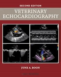
Two-Dimensional and M-Mode Echocardiography for the Small Animal Practitioner
SECOND EDITION
June A. Boon MSInstructor and Echocardiographer College of Veterinary Medicine and Biomedical Sciences Colorado State University Fort Collins, Colorado, USA
This edition first published 2017 © 2017 by John Wiley & Sons, Inc
Editorial offices: 1606 Golden Aspen Drive, Suites 103 and 104, Ames, Iowa 50010, USA The Atrium, Southern Gate, Chichester, West Sussex, PO19 8SQ, UK 9600 Garsington Road, Oxford, OX4 2DQ, UK
For details of our global editorial offices, for customer services and for information about how to apply for permission to reuse the copyright material in this book please see our website at www.wiley.com/wiley-blackwell.
Authorization to photocopy items for internal or personal use, or the internal or personal use of specific clients, is granted by Blackwell Publishing, provided that the base fee is paid directly to the Copyright Clearance Center, 222 Rosewood Drive, Danvers, MA 01923. For those organizations that have been granted a photocopy license by CCC, a separate system of payments has been arranged. The fee codes for users of the Transactional Reporting Service are ISBN-13: 9781119028536
Designations used by companies to distinguish their products are often claimed as trademarks. All brand names and product names used in this book are trade names, service marks, trademarks or registered trademarks of their respective owners. The publisher is not associated with any product or vendor mentioned in this book.
The contents of this work are intended to further general scientific research, understanding, and discussion only and are not intended and should not be relied upon as recommending or promoting a specific method, diagnosis, or treatment by health science practitioners for any particular patient. The publisher and the author make no representations or warranties with respect to the accuracy or completeness of the contents of this work and specifically disclaim all warranties, including without limitation any implied warranties of fitness for a particular purpose. In view of ongoing research, equipment modifications, changes in governmental regulations, and the constant flow of information relating to the use of medicines, equipment, and devices, the reader is urged to review and evaluate the information provided in the package insert or instructions for each medicine, equipment, or device for, among other things, any changes in the instructions or indication of usage and for added warnings and precautions. Readers should consult with a specialist where appropriate. The fact that an organization or Website is referred to in this work as a citation and/or a potential source of further information does not mean that the author or the publisher endorses the information the organization or Website may provide or recommendations it may make. Further, readers should be aware that Internet Websites listed in this work may have changed or disappeared between when this work was written and when it is read. No warranty may be created or extended by any promotional statements for this work. Neither the publisher nor the author shall be liable for any damages arising herefrom.
Library of Congress Cataloging-in-Publication Data
Names: Boon, June A., author.
Title: Two-dimensional and M-mode echocardiography for the small animal practitioner / June A. Boon.
Description: Second edition. | Ames, Iowa : John Wiley & Sons Inc., 2017. | Includes bibliographical references and index.
Identifiers: LCCN 2017015710| ISBN 9781119028536 (pbk.) | ISBN 9781119028550 (Adobe PDF) | ISBN 9781119028567 (epub)
Subjects: LCSH: Veterinary echocardiography. | MESH: Echocardiography–veterinary | Handbooks
Classification: LCC SF811 .B663 2017 | NLM SF 811 | DDC 636.089/61207543–dc23
LC record available at https://lccn.loc.gov/2017015710
A catalogue record for this book is available from the British Library.
Wiley also publishes its books in a variety of electronic formats. Some content that appears in print may not be available in electronic books.
This book is dedicated to my family – you all make my life rich.
CONTENTS
Preface
About the Companion Website
Chapter 1: The Basics
Echocardiographic Applications
Some Indications for an Echocardiogram
Cardiac Anatomy
Orientation of the Heart in the Thorax
How To
Chapter 2: Knobology for the Echocardiogram: Improving Image Quality
Depth
Frequency
Gain
Time gain compensation (TGC)
Grey Map
Dynamic Range
Rejection
Focus
Scan Area
Harmonics
Sweep Speed
Compounding and Cross Beam
Chapter 3: Two-Dimensional Imaging Planes and Subjective Assessment
RIGHT PARASTERNAL IMAGING PLANES: LONG-AXIS VIEWS
Left Ventricular Inflow Outflow (Left Ventricular Outflow)
Four-Chamber
RIGHT PARASTERNAL IMAGING PLANES: SHORT-AXIS (TRANSVERSE) VIEWS
Left Ventricle at the Papillary Muscles
Left Ventricle at the Chordae Tendineae
Left Ventricle at the Mitral valve
Heart Base: At the Left Atrium
Heart Base: At the Pulmonary Artery
LEFT PARASTERNAL IMAGING PLANES: CRANIAL LONG-AXIS VIEWS
Left Ventricular Outflow
Right Atrium and Auricle
Pulmonary Artery
LEFT PARASTERNAL IMAGING PLANES: CRANIAL TRANSVERSE VIEWS
Pulmonary Artery and Tricuspid Valve
Heart Base of the Left Auricle
LEFT PARASTERNAL IMAGING PLANES: APICAL VIEWS
Apical Four-Chamber
Apical Five-Chamber
Chapter 4: Imaging Planes: Technique in the Dog and Cat
RIGHT PARASTERNAL IMAGING PLANES: LONG-AXIS VIEWS
Inflow Outflow (Left Ventricular Outflow)
Four-Chamber
SHORT-AXIS (TRANSVERSE) VIEWS
Technique in the Dog and Cat
LEFT PARASTERNAL IMAGING PLANES: CRANIAL LONG-AXIS VIEWS
Left Ventricular Outflow
Right Atrium and Auricle
Right Ventricular Outflow (Pulmonary Artery)
LEFT PARASTERNAL IMAGING PLANES: TRANSVERSE VIEWS
Transverse Heart Base
Left Auricle
LEFT PARASTERNAL IMAGING PLANES: APICAL VIEWS
Apical Four-Chamber
Apical Five-Chamber
Chapter 5: M-Mode Echocardiography
Principles of M-Mode Echocardiography
M-Mode of the Left Ventricle
M-Mode of the Aorta and Left Atrium
M-Mode of the Mitral Valve
Chapter 6: Measurement and Assessment of Two-Dimensional and M-Mode Images
Measurement of the Left Ventricular Chamber
Measurement of the Aorta and Left Atrium
Measurement of the Mitral Valve
Assessment of Two-Dimensional and M-Mode Measurements
Echocardiographic Reference Values
References: Breed-Specific Echocardiographic Reference Ranges
Chapter 7: Echocardiographic Features of Common Acquired Heart Diseases
Degenerative Mitral Valve Disease
Hypertrophic Cardiomyopathy
Dilated Cardiomyopathy
Unclassified Cardiomyopathy
Pericardial Effusion
Hemangiosarcoma
Aortic Body Tumors
Endocarditis
Recommended Reading
Glossary
Index
EULA
List of Tables
Chapter 6
Table 6.1
Table 6.2
Table 6.3
Table 6.4
Table 6.5
Table 6.6
List of Illustrations
Chapter 1
Figure 1.1
Heart Diagram.
Diagram of the heart showing the chambers, valves and vessels of the heart. Flow through the heart is indicated. Note the relationships between the mitral valve and the aorta, and the pulmonary artery and the tricuspid valve. For details of abbreviations used in the figures, see the Glossary.
Figure 1.2
Canine lateral thoracic radiograph.
This lateral radiograph shows the typical orientation of the dog's heart in the thorax. The triangle superimposed over this radiograph represents the sheet of sound coming from the transducer. Note that the transducer crystals need to be directed to the mid lumbar spine in order to create the long-axis image. Short-axis echocardiographic images of the heart have the sheet of sound oriented 90° to the long axis.
Figure 1.3
Canine lateral thoracic radiograph – deep-chested dog.
This lateral radiograph shows the typical orientation of the dog's heart in the thorax when the dog has a deep chest, such as the Doberman Pinscher, Irish Wolfhound and the German Shepherd. The triangle superimposed over this radiograph represents the sheet of sound coming from the transducer. Because the heart is oriented more vertically in the thorax, the sheet of sound needs to be directed more to the tail than the mid lumbar spine, and the transducer needs to be located more cranially and dorsally to be in front of the heart. Short-axis echocardiographic images of the heart have the sheet of sound oriented 90° to the long axis.
Figure 1.4
Feline lateral thoracic radiograph.
This lateral radiograph shows the typical orientation of the cat's heart in the thorax. Note that it is aligned more parallel to the sternum than the dog's heart. The triangle superimposed over this radiograph represents the sheet of sound coming from the transducer. The sheet of sound needs to be directed more towards the thoracolumbar junction than in the dog in order to create the long-axis image. Short-axis echocardiographic images of the heart have the sheet of sound oriented 90° to the long axis.
Figure 1.5
Shaving on the right side.
On the right side of the thorax in both dogs and cats, shave from behind the front leg to about the sixth intercostal space, from the costochondral junction to the sternum.
Figure 1.6
Shaving on the left side.
On the left side of the thorax in both dogs and cats shave from behind the front leg at the costochondral junction to just past the last rib in a triangular shape. Shaving past the last rib is essential in order to obtain good image quality and long left apical views.
Figure 1.7
The Echocardiographic Scan Table.
(a–c) A table with cut-outs that allow placement of the transducer from below the thorax enhances image quality because it reduces lung interference. Here, three echo scan tables are seen. (a) A homemade table with various cut-outs for different scanning techniques. (b) A commercially available table with a single cut-out that is used for both left and right parasternal imaging. An overlay (line with arrows) with a smaller hole can be positioned over the larger hole in order to accommodate small dogs and cats. (c) Another commercially available scan table with cut-outs at each end of the table, one located at the edge of the table for left parasternal imaging. Many other options exist, but all improve image quality and minimize frustration.
Figure 1.8
The Echocardiographic Scan Table and Animal Placement.
Place the animal in right or left lateral recumbency over the table cut-out. Here, the dog is placed in right lateral recumbency on the table with the thorax located over the cut-out. The dog's head is to the left side of this image. The transducer is positioned against the right side of the thorax from below the dog to obtain the right parasternal imaging planes. The heart drops through the lungs, and image quality is generally improved over imaging that is done with the animal placed in left lateral recumbency and obtaining right parasternal images from above. The same principles apply when obtaining left parasternal images: the animal is place left-side down over the cut-out.
Chapter 2
Figure 2.1
Depth.
(a, b) Adjust depth so that the echo image fills the sector. (a) Wasted space below the image; (b) An appropriate depth setting. For details of abbreviations used in the figures, see the Glossary.
Figure 2.2
Transducer frequency.
(a, b) Higher-frequency transducers provide better image resolution but less depth penetration, while low-frequency transducers can image deeper structures but have poorer resolution. (a) A high-frequency transducer used in a dog. (b) A lower-frequency transducer used in the same dog. Image quality is poorer in panel (b) than in panel (a).
Figure 2.3
Gain.
(a–c) Gain adjusts the overall brightness of the image. Adjust gain to see structures clearly. (a) A low gain setting, where the image is too black and details are difficult to see; (b) A very high gain setting, where the pixels begin to blend together and details are lost. (c) An appropriate gain setting.
Figure 2.4
Time Gain Compensation (TGC) curve.
(a–c) This adjusts for attenuation (loss of sound energy) as the sound beams travel deeper into the body. Using overall gain would increase gain over the entire image, while TCG sliders control gain at specific depths. (a) Far-field attenuation with low gain. The appropriate TGC sliders should be moved to the right in order to increase brightness at that depth; (b) Too little gain in the near field; the top few TGC sliders should be moved to the right in order to increase the level of brightness in the near field; (c) The TGC curve adjusted so that gain levels are similar from near to far field.
Figure 2.5
Grey maps.
(a, b) Grey maps change the intensity of grays in the image. (a) A gray map with more contrast than in panel (b), which shows softer grays. Adjust based on personal preference without losing image detail.
Figure 2.7
Rejection.
(a, b) Low-intensity sound can be “rejected” – removed from the image. (a) An M-mode image without a lot of rejection; (b) The same image with a lot of rejection added; this results in blacker chambers. Here, so much rejection has been added that some details are lost. Rejection is used more often in M-mode imaging than in two-dimensional imaging.
Figure 2.9
Harmonics.
(a, b) Having harmonics on usually provides better resolution and fewer artifacts. It does not always work, however, depending on the transducer frequency and the patient. Turn harmonics on or off and see which setting provides the best image quality. (a) Harmonics on; (b) Harmonics off.
Chapter 3
Figure 3.1
Normal right parasternal left ventricular inflow outflow view in the dog
. (a, b) The ideal long-axis inflow outflow image is horizontal across the sector, has a parallel wall and interventricular septum, has an aorta that shows the aortic valve dash in its middle during diastole, and has a mitral valve that opens well into the left ventricular chamber. (a) In diastole, as seen here, the mitral valve, chamber size, wall thicknesses and left atrial size are assessed. See text for features of the normal inflow outflow view in the dog; (b) In systole, the mitral valve is assessed for prolapse. Here, the mitral valve does not curve backwards into the left atrial chamber. For details of abbreviations used in the figures, see the Glossary.
Figure 3.2
Abnormal right parasternal inflow outflow view.
(a) This diastolic frame shows a ventricular septum that curves upward (arrow), consistent with a dilated left ventricular chamber, a left atrium that is larger than the aorta, consistent with left atrial dilation and an anterior mitral valve leaflet that is thickened; (b) This systolic frame shows prolapse (arrow) of the mitral leaflet into the left atrial chamber.
Figure 3.3
Abnormal right parasternal inflow outflow view.
This image shows right ventricular hypertrophy with a wall thickness that is much more than left ventricular wall thickness. The pressure overload that is creating this hypertrophy causes the interventricular septum to curve downward. A component of this downwardly curved septum may be secondary to decreased preload in the left ventricular chamber. The result is that the left ventricular walls appear thick because of poor volume in the heart. Only about two wall or septal thicknesses fit in this left ventricular chamber. There is a fibrous band of tissue extending down from the interventricular septum into the left ventricular outflow tract (arrow). This is one finding of subaortic stenosis in the dog which may also be creating the hypertrophic appearance in this heart.
Figure 3.4
Abnormal right parasternal inflow outflow view
. This image shows a right ventricular chamber that is much larger than the left ventricular chamber. The right ventricular chamber should be about one-third to one-half the size of the left ventricular chamber.
Figure 3.5
Normal right parasternal left ventricular inflow outflow view in the cat.
The ideal long-axis inflow outflow view image is horizontal across the sector, the septum may bow slightly upward at end-diastole, the left atrium may appear larger than the aorta (it is not larger in this image), the interventricular septum may normally extend down in the left ventricular outflow tract to a small degree (arrow), the interventricular septum and wall thicknesses are similar, about three to four wall or septal thicknesses should fit into the left ventricular chamber at end-diastole, the right ventricular chamber should be one-half or less the size of the left ventricular chamber, the right wall thickness should be about one-half the thickness of the normal left ventricular wall.
Figure 3.6
Abnormal right parasternal left ventricular inflow outflow view in the cat.
(a) This long-axis view shows a hypertrophied interventricular septum and wall – only about two of these thicknesses can fit into the left ventricular chamber at end-diastole, the mitral valve is normal with no irregularities during diastole when it is wide open; (b) This long-axis view shows systolic anterior mitral valve motion (SAM) of the mitral valve (arrow). This should not occur, and is a sign of left ventricular outflow tract obstruction.
Figure 3.7
Normal right parasternal four-chamber view in the dog.
(a) In diastole, the normal four-chamber view should show an interventricular septum and left ventricular wall that are parallel to each other, an interventricular septum that is straight, an interatrial septum that is straight, and mitral valve leaflets that are thin from the base of the leaflets to their tips; (b) The mitral valve leaflets should not prolapse during systole. The closed leaflets should not break the plane defined by the mitral annulus (hinge points of the leaflets). It is normal to see chordae tendineae extend from the leaflets during systole, and this is the reason why irregularities of the valve are not assessed during systole.
Figure 3.8
Normal right parasternal four-chamber view in the cat.
During diastole, the normal four-chamber view should show an interventricular septum and left ventricular wall that are parallel to each other, an interventricular septum that is straight, an interatrial septum that is straight, and mitral valve leaflets that are thin from the base of the leaflets to their tips.
Figure 3.9
Abnormal right parasternal four-chamber view in the dog.
This four-chamber view shows a curved interventricular septum (arrow) consistent with left ventricular dilation, a thick and irregular anterior mitral valve leaflet (arrow), and an interatrial septum that is directed upward on the right side of the sector image consistent with left atrial dilation.
Figure 3.10
Abnormal right parasternal four-chamber view in the dog.
The atrial septum is curved downward in the four-chamber view, consistent with right atrial dilation. The right atrium should be similar in size to the left atrium.
Figure 3.11
Cranial and caudal vena cava.
(a) Lifting the transducer up towards the dog's thorax from the four-chamber view, making the angle between the transducer and the thorax smaller, brings the caudal vena cava into view. This can be mistaken for right atrial dilation. (b) Continuing to lift the transducer up towards the thorax and adding a little rotation will show more of the caudal and the cranial vena cava.
Figure 3.12
Abnormal right parasternal four-chamber view in the dog.
Right ventricular wall thickness should be about one-half the thickness of the left ventricular wall in the normal heart. This heart has right ventricular hypertrophy, with a wall thickness that is much thicker than the left ventricular wall. The interventricular septum is also thicker than the left ventricular wall and is part of the right ventricular hypertrophic pattern.
Figure 3.13
Normal right parasternal transverse left ventricle view in the dog and cat.
The normal transverse left ventricular view should have an outer shape that is circular, a left ventricular chamber that is shaped like a mushroom, papillary muscles that are similar in size, and a right ventricular chamber dimension above the septum that is no more than one-third to one-half the size of the left ventricular dimension. A third papillary muscle is a normal finding in both cats and dogs, and is seen on the feline video.
Figure 3.14
Abnormal right parasternal transverse left ventricle view.
A flattened ventricular septum occurs when the right ventricular chamber is either dilated or hypertrophied secondary to volume or pressure overload. Here, the septum is flattened because of right ventricular volume overload.
Figure 3.15
Abnormal right parasternal transverse left ventricle view.
The right ventricular wall should be no more than one-half the thickness of the left ventricular wall. Right ventricular hypertrophy occurs secondary to pressure overload because of pulmonary hypertension or pulmonic stenosis. Here, the left ventricular wall and septum also appear thick because of poor preload in the left ventricular chamber.
Figure 3.16
Normal right parasternal transverse left ventricle view.
Irregularities (arrows) on the right ventricular side of the interventricular septum are a normal finding. They represent the trabeculae found in the right side of the heart.
Figure 3.17
Normal right parasternal transverse chordae tendineae view.
Bright white lines seen where the papillary muscles used to be represent the chordae tendineae on the transverse view in a cat. This is the level at which left ventricular measurements are made.
Figure 3.18
Normal right parasternal transverse mitral valve view.
Mitral valve leaflets move away from each other during diastole, with the anterior leaflet moving up towards the interventricular septum and the posterior leaflet moving down towards the left ventricular wall. The leaflets move towards each other during systole. This view is often called the “fish mouth” view.
Figure 3.19
Normal right parasternal transverse heart base: aorta and left atrium view.
The aorta is a circular or clover-shaped structure in the middle of the sector image. The junctions of the three aortic valve cusps which are seen are often called the “Mercedes” sign. The left atrium is below the aorta, and to the right side is the left atrial appendage. The aorta and left atrial chamber should appear to be similar in size during the cardiac cycles. Frame-by-frame analysis shows an atrial chamber that is about 1.5-fold larger than the aorta at end-systole, when the atrial size is at its maximum in both cats and dogs. The right side of the heart is seen at the top of the sector image, but subjective assessment of the tricuspid valve and any pulmonary valve that is seen is limited. A pulmonary vein is often seen entering the left atrial chamber (arrow).
Figure 3.20
Abnormal right parasternal transverse heart base: aorta and left atrium view.
A left atrium that is much larger than the aorta is consistent with left atrial dilation. Side-lobe artifact is often seen within the chambers of the heart. Here, the irregular hazy white structures floating within the left atrial chamber is a side-lobe artifact and should not be confused for a thrombus or mass. Always look at other views to confirm abnormalities. A side-lobe artifact will appear different or be absent on other left atrial views.
Figure 3.21
Abnormal right parasternal transverse heart base: aorta and left atrium view.
A soft-tissue structure within the left atrial appendage (arrow) is consistent with a thrombus in cats. This image also shows some pericardial effusion and a large left atrium.
Figure 3.22
Normal right parasternal transverse heart base: aorta and pulmonary artery view.


