9,99 €
Mehr erfahren.
- Herausgeber: tredition
- Kategorie: Fachliteratur
- Sprache: Englisch
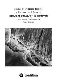
The book presents 573 scanning electron microscopic (SEM) pictures of differently designed, untreated and treated human tooth specimens of molars and incisors in magnifications of more than 50,000 times. The specimens were made from permanent sound, caries-free teeth that were extracted for orthodontic reasons. Sawing and fracture areas were prepared. Treatments were done with phosphoric acid, polyacrylic acid, EDTA, sodium hydroxide and phosphoric acid esters. Many unique pictures show the detailed structures of untreated and treated human enamel and dentin. This collection of SEM pictures might be quite unique in the world. This picture book is ideal for teaching purposes. The pictures are not discussed or interpreted, but it is only explained what is seen.
Das E-Book können Sie in Legimi-Apps oder einer beliebigen App lesen, die das folgende Format unterstützen:
Seitenzahl: 125
Veröffentlichungsjahr: 2021
Ähnliche


Cover picture: SEM picture of phosphoric acid-etched enamel/dentin junction (see PicSer-2 - Pic. 3b)
1st Edition
Imprint
Janda, Ralf, SEM Picture Book of Untreated & Treated Human Enamel & Dentin
Publisher: tredition GmbH, Halenreie 40-44, D-22359 Hamburg
www.tredition.de
Copyright © 2021 by Janda, Ralf
Cover: Janda, Ralf
All rights reserved. No part of this book may be reproduced or transmitted in any form or by any means without the written permission of the copyright holder.
ISBN 978-3-347-35177-6 (e-Book)
In Memoriam
Queeny, Buffy & Vinny
Welcome
Shawny & Lenny
Contents
Contents
Preface 1st Edition / 2nd Version
Preface - 1st Edition
Introduction
Preparation of the Specimens
1 Design
2 Mechanical Treatment
2 Chemical Treatment
3 Dehydration and Drying Methods
4 Replica Technique
5 Picture Series
Picture Series 1
Picture Series 2
Picture Series 3
Picture Series 4
Picture Series 5
Picture Series 6
Picture Series 7
Picture Series 8
Picture Series 9
Picture Series 10
Picture Series 11
Picture Series 12
Picture Series 13
Picture Series 14
Picture Series 15
Picture Series 16
Picture Series 17
Picture Series 18a
Picture Series 18b
Picture Series 19a
Picture Series 19b
Picture Series 20a
Picture Series 20b
Picture Series 21a
Picture Series 21b
Picture Series 21c
Picture Series 22a
Picture Series 22b
Literature
Curriculum Vitae
Preface 1st Edition / 2nd Version
Editorial revisions, corrections of write errors, and an optimization of the bibliography were done in the 2nd version of this e-Book.
Best regards, Ralf
December 2023
Preface - 1st Edition
Dear readership,
this is a very personal book. The shown SEM pictures were made between 1985 and 1989 during the research work for my habilitation. Since then much has changed, but it can be taken for granted that the structures of human enamel and dentin remained unchanged. Therefore, the shown pictures are certainly still relevant and will remain so in future. Only few of the presented SEM photos were published earlier so that it truly makes sense to scan all the 573 photos to present the different appearances of untreated and treated human enamel and dentin. You can find pictures with magnifications higher than 50,000 times.
I do not discuss or interpret the pictures but only explain what is seen. Therefore, enjoy the pictures and see what nature has developed and built.
Best regards
Ralf
Falkensee, June 2021
Introduction
This book shows 573 SEM pictures of differently designed untreated and differently chemically treated human tooth specimens of molars and incisors. The pictures are not discussed or interpreted, but it is only explained what is seen. The specimens are made from permanent sound, caries-free teeth that were extracted because of orthodontic reasons. The teeth were stored in 0.1 mass% aqueous thymol solution at room temperature until usage.
After preparation, dehydration, and drying, all specimens were gold-sputtered and investigated with the following scanning electron microscopes
- Type JSM-23, Jeol Ltd., Tokyo, JP
- Stereoscan 150 MK 2 and Stereoscan 100, Cambridge Ltd., Cambridge, GB
I must especially thank the Engineer of Photography Mr. H. Everts for realizing the exceptional SEM photographs and Degussa AG, Hanau, Germany (now: Evonik AG, Essen, Germany) for providing the devices and the equipment.
Some SEM pictures were already published in the articles [1-5]. The following pictures:
PicSer-2: 3a, b, c /15/7a, b, c/8a, b, c/11a
PicSer-4: 2a, b, c/4d/ 5a, b, c, d, e
PicSer-5: 2a, b/3d/15c/18/18a/21a
PicSer-7: 5b/13/14c
PicSer-21a: 3b/4a, b, c/5/6/7
PicSer-21b: 1a/3b
PicSer-22a: 3b, c
are taken from my article [5] and are shown in this book with permission of Elsevier Copyright Clearing Center, Danvers, MA, USA. This article is available online from the Biomaterials website.
Other pictures were already published in my articles [1-3]. The editor Phillip Verlag, München, Germany does not exist anymore. These publications are not available online, but upon request I can provide PDF files free of charge.
For more detailed information about the preparation and treatment methods or about the interpretation of the pictures, I kindly refer to my publications [4, 5] that are available online.
Please note: For a fee, you can buy JPG or TIFF files up to 600 dpi for different purposes. Upon request, please mail to [email protected] to get a price list.
Preparation of the Specimens
1 Design
The specimen designs were made as described in the following paragraphs.
Design type 1 (DesTyp-1): Vertical and horizontal saw-cuts were made in molar teeth, resulting in a step-like design (Fig. 1). Therefore, it was possible to observe the parallel and perpendicular course of enamel prisms and dentin tubules. Since only one half of the tooth specimen was treated, it was possible to investigate the same area untreated and treated.
Fig. 1: Design type 1 (step-like design).
Design type 2 (DesTyp-2): Vertical and horizontal saw-cuts were made in molar teeth resulting in a step-like design followed by a fracture process from cervical to occlusal along the tooth axis done with a strong and sharp knife (Fig. 2). The knife was placed cervically in order not to damage the occlusal surface. In the first case, the center of the step- like sawn tooth specimen was treated before the fracture process so that treated sawing areas adjacent to untreated fracture areas could be investigated. In the second case, the fracture area of the second half of the fractured tooth specimen was treated so that it was possible to investigate treated sawing areas adjacent to treated fracture areas.
Fig. 2: Design type 2 (step-like design followed by a fracture process).
Design type 3 (DesTyp-3): Discs of 2 to 3 mm thickness perpendicular to the tooth axis were prepared from molar teeth by sawing.
Designs for chemical treatment (DesTyp-3a)
To prepare the specimens for PicSer-11 to 19b, the discs were fractured into two halves using a very sharp knife. Each of the two halves was treated by completely immersing in the respective treatment medium and then SEM-investigated.
Designs for replica technique (DesTyp-3b)
The disk specimens for PicSer-21a to 22b were differently treated as described in chapter “Replica Technique”.
Design type 4 (DesTyp-4): Incisors were sectioned by a horizontal saw, cut into two halves.
2 Mechanical Treatment
The molar teeth of DesTyp-1 and DesTyp-2 were mounted on MMA/PMMA resin (Technovit 4004, Kulzer GmbH, Wehrheim, Germany) so that none of the investigated areas was touched with the resin. Before the SEM investigation, the resin base was cut off.
The teeth of DesTyp-3 and DesTyp-4 were totally embedded in the aforesaid resin. Sawing of all specimens was done with a diamond low speed saw under rinsing with water (Isomet Low Speed Saw, Bühler-Met GmbH, Esslingen, Germany). After sawing, all sawing areas were polished to high gloss under rinsing with water on a cotton cloth using highly dispersed aluminum oxide (max. particle size 0.05 µm, Bühler-Met GmbH) on a rotating plane disc (Ecomet III, Bühler-Met GmbH).
After sawing and polishing, the specimens were spotless with dist. water for 5 min in an ultrasonic bath to remove all residues of the polishing medium.
Before ultrasound cleaning energy dispersive X-ray analysis founds Al-signals deriving from the aluminum oxide polishing medium, what was not the case afterward anymore. SEM investigations did not show aluminum oxide particles on the tooth surfaces, and the tubules were not smeared up. However, very seldom some few residues of the polishing and embedding material were found in the tubule openings (see PicSer-1).
2 Chemical Treatment
Regarding DesTyp-1, DesTyp-2 and DesTyp-3b, the treatment medium was applied with a little brush. In case that only one part of the specimen was treated, the treatment was done cautiously so that all the other parts remained untouched.
Regarding DesTyp-3a, the fractured specimen was treated by totally immersing in the treatment medium.
The following chemical treatments were done:
Chemical treatment 1 (ChemTreat-1): Treatment of the specimen with 35 mass% aqueous ortho-phosphoric acid (Sigma-Aldrich Chemie GmbH, Taufkirchen Germany) for 3 min. Then rinsing with dist. water for 1 min, followed by different dehydration/drying methods. Finally, gold sputtering was done. The different design types were treated as follows:
a) DesTyp-1: Half-side treatment of the specimen’s sawing areas.
b) DesTyp-2: Half-side treatment of the specimen’s sawing areas and the middle part of the outside enamel surface, but the fracture area remained untreated.
c) DesTyp-2: Half-side treatment of the specimen’s sawing areas, the middle part of the outside enamel surface and only partial treatment of the dentin fracture area (horizontal dentin fracture area).
d) DesTyp-3b: Half-side treatment of the specimen’s sawing and fracture areas (see T-Rep-3).
Chemical treatment 2 (ChemTreat-2): Complete treatment of the DesTyp-3a fragment with 35 mass% aqueous ortho-phosphoric acid (Sigma-Aldrich Chemie GmbH) for 30 min. Then rinsing with dist. water for 5 min, followed by dehydration/drying and gold sputtering.
Chemical treatment 3 (ChemTreat-3): Complete treatment of the DesTyp-3a fragment with 1.2 mol aqueous sodium hydroxide (Sigma-Aldrich Chemie GmbH) (pH 13.5) for 60 min. Then rinsing with dist. water for 5 min, followed by dehydration/drying and gold sputtering.
Chemical treatment 4 (ChemTreat-4): Complete treatment of the DesTyp-3a fragment with an aqueous solution of 0.5 mol of the disodium salt of EDTA (ethylene diamine tetraacetic acid, Sigma-Aldrich Chemie GmbH) adjusted with 1 mol aqueous sodium hydroxide to pH 13.1 for 3 min. Then rinsing with dist. water for 3 min, followed by dehydration/drying and gold sputtering. At high pH values, the disodium salt of EDTA provides its optimal complexing effect.
Chemical treatment 5 (ChemTreat-5): Complete treatment of the DesTyp-3a fragment with an aqueous solution of 0.5 mol of the disodium salt of EDTA (ethylene diamine tetraacetic acid, Sigma-Aldrich Chemie GmbH) adjusted with 1 mol aqueous sodium hydroxide to pH 13.1 for 60 min. Then rinsing with dist. water for 5 min, followed by dehydration/drying and gold sputtering. At high pH values, the disodium salt of EDTA provides its optimal complexing effect.
Chemical treatment 6 (ChemTreat-6): Complete treatment of the DesTyp-3a fragment with an aqueous solution of 10 mass% polyacrylic acid (Degapas 8105S, batch-no TPR 186, Degussa AG, Hanau, Germany) with pH 2.0 for 3 min. Then rinsing with dist. water for 1 min, followed by dehydration/drying and gold sputtering.
Chemical Treatment-7 (ChemTreat-7): Complete treatment of the DesTyp-3a fragment with an aqueous solution of 10 mass% polyacrylic acid (Degapas 8105S, batch-no TPR 186) with pH 2.0 for 60 min. Then rinsing with dist. water for 1 min, followed by dehydration/drying and gold sputtering.
Chemical treatment-8 (ChemTreat-8): Complete treatment of the DesTyp-3a fragment with an aqueous solution of 10 mass% polyacrylic acid (Degapas 8105S, batch-no TPR 186) adjusted with 1 mol aqueous sodium hydroxide to pH 13.3 for 60 min. Then rinsing with dist. water for 5 min, followed by dehydration/drying and gold sputtering.
Chemical treatment 9 (ChemTreat-9): Complete treatment of the DesTyp-3a fragment with undiluted bis(methacryloyloxyethyl) hydrogen phosphate (P-acid-ester, laboratory product) for 3 min. Then rinsing with dist. water for 1 min, followed by dehydration/drying and gold sputtering. Such phosphate compounds were and are still used as adhesion promoters in enamel/dentin adhesives [5, 6].
Chemical treatment 10 (ChemTreat-10): Complete treatment of the DesTyp-3a fragment with an emulsion of equal parts P-acid-ester and dist. water (obtained pH 0.9) for 3 min. Then rinsing with dist. water for 1 min, followed by dehydration/drying and gold sputtering. This was done to investigate if demineralization is stronger in aqueous surrounding.
Chemical treatment 11 (ChemTreat-11): Complete treatment of the DesTyp-3a fragment with a 1:1 mixture of undiluted P-acid-ester and tetra(methacryloyloxyethyl) pyrophosphate (Pyro-ester, laboratory product) for 3 min. Then rinsing with dist. water for 1 min, followed by dehydration/drying and gold sputtering. Such phosphate compounds were used as adhesion promoters in enamel/dentin adhesives [5].
Chemical treatment 12 (ChemTreat-12): Complete treatment of the DesTyp-3a fragment with an emulsion of equal parts of the 1:1 mixture of P-acid-ester and Pyro-ester (obtained pH 1.4) for 3 min. Then rinsing with dist. water for 1 min, followed by dehydration/drying and gold sputtering. This was done to investigate if demineralization is stronger in aqueous surrounding.
Fig. 3: Structural formula of bis tetra(methacryloyloxyethyl) pyrophosphate) hydrogen phosphate (P- acid-ester).
Fig. 4: Structural formula of tetra(methacryloyloxyethyl) pyrophosphate (Pyro-ester).
3 Dehydration and Drying Methods
The direct or indirect method can be used to investigate natural teeth with the scanning electron microscope (SEM) (Fig. 5) [4]. In case the direct method is chosen, the method of dehydration and drying is of essential importance to avoid shrinkage artifacts.
The following dehydration and drying methods were investigated:
Dehydration/drying method 1 (DeDry-1): Vacuum desiccation (glass filter pump vacuum) for 48 h over silica gel.
Dehydration/drying method 2 (DeDry-2): Vacuum desiccation (glass filter pump vacuum) for 72 h over silica gel, followed by 72 h over phosphorous pentoxide.
Dehydration/drying method 3 (DeDry-3): Dehydration in water/ethanol with increasing concentrations of 70, 80 and 96 % ethanol for 24 h at each step and finally three times in fresh 100 % ethanol for 24 h. Then vacuum desiccation (oil pump vacuum) over phosphorous pentoxide for 24 h.
Dehydration/drying method 4 (DeDry-4): Dehydration in water/ethanol with increasing concentrations of 70, 80 and 96 % ethanol for 24 h at each step and finally three times in fresh 100 % ethanol for 24 h. Then vacuum desiccation (oil pump vacuum) without drying medium for 2 h.
Dehydration/drying method 5 (DeDry-5): Dehydration in water/ethanol with increasing concentrations of 70, 80 and 96 % ethanol for 24 h at each step and finally three times in 100 % ethanol for 24 h. Further dehydration in ethanol/acetone with increasing concentrations of 80 % and 96 % acetone at each step for 24 h and finally three times in fresh 100 % acetone for 24 h each time. Then vacuum desiccation (oil pump vacuum) without drying medium for 2 h.
Dehydration/drying method 6 (DeDry-6): Dehydration in water/ethanol with increasing concentrations of 70, 80 and 96 % ethanol for 24 h at each step and finally three times in fresh 100 % ethanol for 24 h. Further dehydration in ethanol/acetone with increasing concentrations of 80 % and 96 % acetone at each step for 24 h and finally three times in fresh 100 % acetone for 24 h. Then vacuum desiccation (oil pump vacuum) without drying medium for 10 min. Then again three times, storage in fresh 100 % acetone for 24 h. Vacuum desiccation (oil pump vacuum) was done after each of the final three acetone steps without drying medium for 10 min. The intent was to discover whether further water removal was possible or not.
Dehydration/drying method 7 (DeDry-7): Dehydration in water/ethanol with increasing concentrations of 70, 80 and 96 % ethanol for 24 h at each step and finally three times in fresh 100 % ethanol for 24 h. Further dehydration in ethanol/acetone with increasing concentrations of 80 % and 96 % acetone at each step for 24 h and finally three times in fresh 100 % acetone for 24 h. Then, the critical point drying (CPT) by flushing three times with fresh carbon dioxide was performed. The last flush was left to evaporate within 1 h. Gold sputtering was done immediately afterward. Further hints and literature about CPT is given by [4, 5]
Conclusions
DeDry-7 produced the best results. The fewest artifacts were found, and particularly the fine structures were perfect visible. Almost the same quality level was achieved with DeDry-5 and DeDry-6. No differences were observed between DeDry-5 and DeDry-6. Some more drying artifacts and less visible fine structure were fond for DeDry-3 and DeDry-4 but nearly no differences were found between these methods. The worst results were produced by methods DeDry-1 and DeDry-2. Therefore, it was concluded to use DeDry-5 as the standard method.
4 Replica Technique


