20,99 €
Mehr erfahren.
- Herausgeber: Crowood
- Kategorie: Geisteswissenschaft
- Sprache: Englisch
Extreme macrophotography opens up a new world for photographers, particularly biologists. By photographing subjects way beyond just life size magnifications, this book takes you a step closer to the very cells that make up organisms. Written with clarity and detail, Julian Cremona's book is the perfect guide and sister title to Extreme Close-Up Photography and Focus Stacking. His enthusiasm for and knowledge of his subject makes this book an essential companion for everyone interested in photography and discovering the minutiae of the natural world.
Das E-Book können Sie in Legimi-Apps oder einer beliebigen App lesen, die das folgende Format unterstützen:
Veröffentlichungsjahr: 2018
Ähnliche


Beyond Extreme Close-Up Photography
Julian Cremona
CROWOOD
First published in 2018 by
The Crowood Press Ltd
Ramsbury, Marlborough
Wiltshire SN8 2HR
www.crowood.com
This e-book first published in 2018
© Julian Cremona 2018
All rights reserved. This e-book is copyright material and must not be copied, reproduced, transferred, distributed, leased, licensed or publicly performed or used in any way except as specifically permitted in writing by the publishers, as allowed under the terms and conditions under which it was purchased or as strictly permitted by applicable copyright law. Any unauthorised distribution or use of thistext may be a direct infringement of the author’s and publisher’s rights, and those responsible may be liable in law accordingly.
British Library Cataloguing-in-Publication Data
A catalogue record for this book is available from the British Library.
ISBN 978 1 78500 466 7
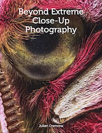
Cover Photograph: Eye of the small elephant hawkmoth. Forty images have been focus stacked to create this composite image using Helicon Focus software. (×6 magnification, Canon 7D mk2, 65mm MPE lens ƒ5.6 ISO 100, with diffuse twin macro flash.) through several rinses in clean water to remove detritus.
Frontispiece: Part of the underside of a rose chafer beetle, Cetonia aurata. (×7 magnification, Canon 7D mk2 with 65mm MPE, 32mm of extension tube, ƒ5.6 ISO 100 twin macro flash, composite of thirty-two images.)
CONTENTS
Preface
1What is Magnification?
2What Cameras are Best?
3Techniques to go Beyond Macro
4An Introduction to Microscopes
5Lighting Extreme Close-ups
6Support and Preparation
7Focus Stacking
8Inspiration for Extreme Close-up Photos
References and Further Reading
Index
Preface
‘The contribution of visible life to biodiversity is very small indeed.’
Sean Nee writing in Nature, 2004.
Iam a regular reader of the photographic press. The articles I enthusiastically lap up are as diverse as possible because this is one of my pet loves; taking photographs. My grandchildren know, just like their parents, that a camera will always be there to record the moment. Any moment of their lives. One, which some find strange, is the appearance and disappearance of teeth as they grow up; these can present technical challenges along with the personal level of dealing with my model. This should not come as a surprise to people who know me, because they realize that my passion is for anything natural and biological. The primary purpose of my travels around the globe is in the search for wilderness and then to record, through my photography, the life that exists there no matter what the size or type of organism. Learning through this form of research, I am better able to understand the dynamics of the natural world and go on to help in conservation and education.
Spanish scorpion photographed at night with ‘black light’ (365nm wavelength), invisible to human eyes. In daylight the normal colour is yellow. Although this image is not an extreme close-up, it demonstrates that by photographing animals and plants in UV or infrared, some species become more striking and visible to us.
This leads me to a conundrum. I have met people who just photograph flowers (waiting for the insects to disappear first). There is the bird photographer: I was in a very small boat on a river in a remote part of Costa Rica with a guide. I was sharing this with a German photographer, tripod and very long lens at the ready. An exotic bird appeared and we both got snapping. A howler monkey in the tree above; crocodile up close baring its teeth in the water; a bright pink dragonfly at rest on the reed; only my camera was in action. The bird photographer would hardly look.
Now back to the photographic magazines.
I look at the wonderful wildlife photos. My interpretation of wildlife seems to be very different to others. Mammals like foxes, badgers, deer, elephants and tigers grace the pages along with birds and flowers. But surely flowers are not wildlife, are they? Or insects and sea anemones in a tide pool? Of course they are! I, and most biologists, fail to categorize creatures like the press because to me, all life that is wild should include all nature without exception. And here we reach the real ambiguity, because the vast majority of life on Earth is not visible to the naked eye. The photos we typically see in magazines, over and over, are just the tip of a huge biodiversity iceberg and 99.9 per cent of wildlife photographers are only taking photos of that tip. The enormity of what is left is largely overlooked.
Some people will discover more as they use a macro lens to take photos of butterflies and other insects (even macro is seen as a different genre to wildlife in many magazines). There are a host of books and websites out there describing macro techniques, including ‘extreme macro’, but I am amazed that the sites are full of bees and flies. Where are the rest, such as beetle life in a pond or crabs on the seashore? There is so much more out there and still I am talking about the visible creatures, because almost all the biodiversity of life on our planet is not visible to our naked eye. Much of this has never been photographed and it will be in your garden, parkland or nearest wood or pond. Your range of subjects is vast and in practical terms inexhaustible.
The marine pseudoscorpion, Neobisium, photographed in 1998 with a Fuji bridge camera and an SLR lens reversed on to the front and coupled. (×6 magnification, live specimen in seawater, natural light.)
Twenty years ago I photographed a tiny creature called a marine pseudoscorpion on the rocky shores of west Wales. A close relative of the true scorpion, it is a few millimetres long and while being quite common, is rarely seen because of its size. I struggled to find it but on this occasion, I took a photo on a Fuji bridge camera and for the ×4 or ×5 magnification, I had a 50mm SLR lens reversed over the front (a technique called coupling). Until very recently if you Googled this little arachnid, only my rather soft photo appeared as I had it on an educational website. What this illustrates is that we are passing a vast array of species every second of the day, but very few photographers are taking advantage of that bonanza. Instead, like landscape photographers travelling to the classic honeypot sites, there are the nature photographers imaging the ‘big stuff’. I am not complaining as I do the same.
Part of my life-long career in biology has been to educate children across the world in conservation issues. Once I stood up in a Singaporean auditorium and talked to a thousand teenagers, asking them, ‘What is the point of a panda? Given the choice, would you conserve the panda or a bluebottle fly?’, daring them to understand that if we lost pandas the world would carry on; a very sad and genetically poor world, but one that would survive. You can imagine the uproar. Flies are not cuddly creatures and so do not tug at the conservation heartstrings. We need flies to clear up our world of the dead and decaying, not to mention the mountains of dung produced. For the sake of conservation I believe we should be highlighting the wildlife that is so hidden. Entering the realm of the micro-world is such a cliché but it is also very true that it is an exciting, alternative world which so few people ever get to see, let alone photograph. The problem is down to the difficulty of taking photos of very small things and I have tried here and in previous books to redress this problem.
To that end, in 2014 Crowood Press published my first book in the ‘Art and Techniques’ series, Extreme Close-up Photography and Focus Stacking. Discussing everything macro, it covered the area between around a fifth life-size (1:5) down to ×5 magnification (5:1). In 2017, Crowood published An Introduction to Digital Photomicrography by Brian Matsumoto and Carol Roullard. In contrast, this book covers photography of nature with magnifications well in excess of ×100, where the microscope is really the only tool available. I am writing this book to fit between these two and inevitably there will be a small degree of overlap. Starting beyond macro, we are aiming at the rather difficult ×5 (5:1) to ×30 (30:1) magnification region, although the book will in effect be covering anything between life size up to ×100.
Some of the techniques will unavoidably have been mentioned in my other book but I will be taking these forward into new ideas. Most importantly, low cost and DIY systems will be emphasized as much as possible. Some people like categories and names and plenty are bandied about, often incorrectly, like macro photography – which is life size, 1:1. True photomacrography is the name we give to employing optics that are designed for use without an eyepiece on extension tubes or bellows. This can produce the magnifications of up to 40:1 or higher. Photomicrography is the term reserved for photography that requires a compound microscope using an eyepiece connected through a tube to an objective lens at the other end, where the subject is located. The magnifications will overlap, as it can start around 10:1, rising to 2,000:1 (considerably outside the realms of this book).
In fact, the first chapter tackles the very issue of what we mean by magnification. Subsequent chapters consider cameras, techniques and equipment, lighting, supporting methods, focus stacking and at the end, a series of suggestions to try and develop your extreme close-up ideas. In particular, that last chapter helps consolidate some of the detail of the book by bringing the theory into practice with hopefully inspiration on subjects you might never have considered.
While there are only around 500 species of marine fungi across the oceans, these tiny organisms are vital for recycling. The strands of a seashore fungus are growing on a red seaweed, possibly the first photograph ever taken of this species but as so little is known about them, even identification is difficult. (×8 magnification, composite of twenty images taken in seawater.)
I run workshops and during this time, I seem to have developed a number of nicknames based on my extensive use of Blu Tack (other varieties are available, like white tack) and gaffer tape for holding everything together. You can never have enough! Over the years I am sure the people I have met on these workshops and at Quekett Microscopical Club meetings have taught me much more than I have taught them. Two from the club have allowed me to use a few of their amazing photos in this book, namely Mike Crutchley (who, living nearby, has many of his ideas borrowed and used in this book) and Mark Papp. The inspiration from being a member of the Quekett is quite overwhelming and I must thank Phil Greaves, Chris Ramon, Carel Sartory, Ray Sloss, David Spears and Spike Walker for their invaluable help over the years. Phil has been good enough to read through a draft of the book and I thank him for his comments, but the mistakes are entirely my own. My wife Brenda is always there with support and ideas – thank you.
Remember, there are more micro subjects waiting to be photographed in the square mile around your house than you can probably photograph in your lifetime. You just need a hand lens and time to stop and look carefully. Then attempt a photograph and the chances are you are the only person to have done that.
Chapter 1
What is Magnification?
The answer to this question may seem obvious and not that many years ago, it was even more simple to respond. Sherlock Holmes would have reached in his pocket and retrieved a large magnifying glass and the object would double in size to make it clearer to see. But today with a plethora of digital devices, the explanation has become distorted and more difficult to interpret.
Fig. 1.1
Head of an opossum shrimp, photographed live in seawater with dark field lighting. Common in estuaries and on the shore, the family of mysids to which it belongs are believed to be a primitive group of crustaceans. (×4 magnification, Canon 7D mk2 with 65mm MPE lens at ƒ5.6, composite of 25-image stack.)
WHAT DO WE MEAN BY MAGNIFICATION?
With so much nutritional information needing to be put on food labels these days, manufacturers will typically make the font size small, sometimes so small it is almost impossible to read. As we age, the ability to resolve detail with our eyes can deteriorate, particularly in low light. Perhaps it is a feature of our modern age that legal information at the bottom of documents is in a tiny font, so we ignore it. Few people carry a large magnifying glass but invariably a smartphone will be in the pocket or bag. A quick snapshot of the label or document appears on the screen and with a pinch or two of your fingers, the image is magnified so the text can now be read.
Clearly, this is one way in which we can magnify a subject. With a large enough screen, we can blow up our photos so that an image of our dog is so enlarged we might make out a tick on the side of the body. We could crop the image so that we just have the small part we want and then on the screen, it appears that we have a highly magnified subject. To produce anything vaguely useful, this technique relies on a high number of pixels being present in the original photograph as cropping the photograph possibly removes many millions of pixels.
Fig. 1.2
Green hairstreak butterfly photographed on a gorse flower. The photo is true macro 1:1 as the width of the image measures 35mm and in real life, the subject matter was 35mm across.
The green hairstreak butterfly in Fig. 1.2 is an example. The image is an 18-megapixel photo and to enlarge the head, this was cropped with the resulting photo becoming just 709 by 476 pixels, which is 337,484 pixels in total, making a 0.33 megapixel image. With so few pixels in the photo, it would appear very small on a screen and this can be improved by boosting the number, in this case to 2,500 by 1,678, around 4 megapixels. This can be done in any photo editor, followed by further enhancement to reduce any noise and increasing clarity and sharpening. It is printed small on this page to show that despite a reduction in quality, it may be acceptable.
Fig. 1.3
A crop of just the head of the green hairstreak in Fig. 1.2, showing it considerably enlarged but the image is of a significantly lower quality.
The head of the butterfly may be magnified, but by how much? The original photograph covers an area approximately 35mm across and we could print a scale to indicate the size of the butterfly. This is usual in scientific literature but not always appropriate elsewhere. The cropped image is 3.5mm across and so if the images are printed to the same size, the enlarged section has been magnified by ×10. But this is still rather vague and cropping is not the best way to magnify a subject and should be used when no other technique is available. If we wish to give a specific figure to magnification there has to be a better way.
The conventional method harks back to film, in particular 35mm film. The green hairstreak butterfly would be classified as a life-size image or macro photograph. If the photo had been made on film, after processing, the size of the actual butterfly would be identical to the outline of the image on the film. You could lay one on top of the other and they would be the same. The field of view of the original is 35mm across, the same as in film. This true macro is given the ratio of 1:1, referring to the size on the film to the size of the subject in real life. If the photographer of this butterfly could have managed to get closer to the insect and fill the frame so that the back of the wing was touching the left side of the frame and the front leg was on the right frame edge (approximately 18mm across), the butterfly has now doubled in size and the ratio would be 2:1 as the image is now ×2; twice life size. The image could be enlarged on the screen or cropped to create what appears as a higher magnification but for this book, as is generally accepted, we will use the original photograph ratio as a measurement of magnification.
If your photography is hovering around macro using a typical macro or close-up lens then working out the ratio will not be difficult. In fact most of the commonly available lenses will have these marked on the side of the lens barrel, even close-focusing zoom lenses. However, this book aims at significantly higher magnification and for any hope of accuracy in measuring this, you will need to photograph a sharply defined millimetre scale. A standard ruler will not do and unfortunately the desired scale can be quite expensive to buy. What is required is a glass microscope slide, etched with calibration down to 0.01mm. They are sold as a microscope stage micrometer scale and widely available. The stage refers to the fact that it is a glass slide to place on the microscope stage, not to be confused with a graticule that goes inside the eyepiece of a microscope for direct measurement of subjects as you look through the eyepiece at the specimen. Graticules are appreciably more expensive as well.
Fig. 1.4
Stage micrometer slide photographed to show a centimetre scale divided up so that the smallest divisions are 0.1mm (100 microns) across.
Fig. 1.5
Eye of a solitary bee and part of the antenna, photographed with Canon 7D mk2 fitted with bellows and objective lens.
Fig. 1.6
A reference photo of a stage micrometer scale is placed along the edge of the bee image, using the same equipment and set-up as the photo in Fig. 1.5 so that the magnification can be obtained. The red bar represents 0.1mm and can be added as a scale in scientific publications, but has not been included in subsequent photos in this book.
Stage micrometers vary, but the most useful is the glass slide with three separate scales: 1mm, 0.1mm and 0.01mm. The latter is for higher microscope magnification and probably of least use for us here. When you are working with the different methods outlined in the book, for example, photographing the eye of a fly, as well as taking the insect, make sure you have created a reference photo. This reference is of the micrometer scale at that magnification. This only needs to be done once, labelled with the technique and kept safely in a separate folder. Then, if a photo is taken using a particular set-up and you want the magnification, just match up the reference along the edge of the photo, as in Fig. 1.6.
In the case of the bee’s eye a measurement of the length could be made (1.6mm). To calculate the magnification based on the photo:subject ratio, 35mm is divided by the reference scale of 1.73mm, approximately 20:1, which can be represented as ×20.
THE SIGNIFICANCE OF CAMERA SENSORS
As we will see in the next chapter, the digital cameras vary considerably from one to another. There are only a few manufacturers of the sensors and camera producers generate different software (called ‘firmware’) for their cameras, which controls and determines the quality of the output. Unless you intend to crop heavily, the number of sensor pixels is less important than how big it is. Small sensors with large numbers of photosites (the areas that receive light and create the pixels) will be so tightly packed that they can cause interference that manifests itself as noise on the image: distracting random coloured pixels. Any size sensor can be used to produce high magnification, as we will see, and small sensors produce more depth of field (areas of detail that appear in focus).Sensor size seems to influence the magnification although this can be a rather abstract concept. Small sensors, like those in tiny compacts, can create what appears to be substantial magnification.
Our discussion of magnification so far is related to 35mm film. A digital sensor, this size is found in a full frame camera and when lenses – such as a 100mm macro – are fitted to the body the focal length applies, that is, it will be 100mm. APS-C sensors are slightly smaller and the 35mm equivalent for the lenses will not be the same. Owners of Nikon and Sony cameras will be aware that they have to multiply by a crop factor of ×1.5 to identify the correct focal length while Canon APS-C users correct with a factor of ×1.6. The 100mm macro now becomes 150mm and 160mm, respectively. What is usually forgotten in this mental conversion is the value of the APS-C to magnification. With the macro set at 1:1, try photographing a millimetre scale. The full frame model will have a width of 35mm as expected (life size) but the APS-C models will be much less: between 22–24mm. Depending on the camera, divide 35mm by the crop factor, for example, Canon with ×1.6 is 21.9mm.
Fig. 1.7
Donacia beetle. A single image taken at ×7 magnification shows a limited amount of visible detail or depth of field.
Fig. 1.8
Donacia beetle. This is a composite of thirty-five images combined to create a huge depth of field. This is the process of focus stacking.


