79,99 €
Mehr erfahren.
- Herausgeber: Thieme
- Kategorie: Fachliteratur
- Sprache: Englisch
Best of visceral manipulation -- key concepts at a glance
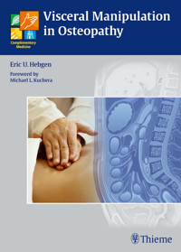
Written by one of the best-known European osteopaths, Visceral Manipulation in Osteopathy explains how to successfully apply the four most common approaches for the manual treatment of abdominal disorders.
The first section provides an overview of the basic principles and techniques of diagnosis and treatment from the greats of osteopathy: visceral manipulation according to Barral; fascial treatment of the organs according to Finet and Williame; circulatory techniques according to Kuchera; and reflex point treatment according to Chapman. Organized by the individual organs, the second section contains "action" photographs that demonstrate osteopathic tests and treatment techniques, plus in-depth information on anatomy, physiology, and pathology.
Features:
- Over 160 clear, marked-up "action" photographs illustrate the different techniques
- Additional graphics display the macroscopic anatomy and topography of the organs
- Practical tips and notes are highlighted throughout for rapid access and quick review
- Organ-tooth interrelationships are demonstrated
Practitioners of osteopathy, physical therapy, or chiropractic will refer to this indispensable clinical guide on a daily basis. The book's breadth and clarity also make it ideal as a textbook for students of visceral manipulation.
Das E-Book können Sie in Legimi-Apps oder einer beliebigen App lesen, die das folgende Format unterstützen:
Seitenzahl: 391
Veröffentlichungsjahr: 2010
Ähnliche


Library of Congress Cataloging-in-Publication Data is available from the publisher.
This book is an authorized translation of the 3rd German edition published and copyrighted 2008 by Hippokrates Verlag, Stuttgart. Title of the German edition: Viszeralosteopathie – Grundlagen und Techniken.
Translator: Sabine Wilms, PhD, Taos, NM, USA
Illustrator: Christiane von Solodkoff, Neckargmünd, Germany
© 2011 Georg Thieme Verlag,Rüdigerstrasse 14, 70469 Stuttgart, Germanyhttp://www.thieme.deThieme New York, 333 Seventh Avenue,New York, NY 10001, USAhttp://www.thieme.com
Cover design: Thieme Publishing GroupTypesetting by Sommer Druck, Feuchtwangen, GermanyPrinted in Germany by Mohn Media, Gütersloh
ISBN 978-3-131-47201-4 1 2 3 4 5 6
Important note: Medicine is an ever-changing science undergoing continual development. Research and clinical experience are continually expanding our knowledge, in particular our knowledge of proper treatment and drug therapy. Insofar as this book mentions any dosage or application, readers may rest assured that the authors, editors, and publishers have made every effort to ensure that such references are in accordance with the state of knowledge at the time of production of the book.
Nevertheless, this does not involve, imply, or express any guarantee or responsibility on the part of the publishers in respect to any dosage instructions and forms of applications stated in the book. Every user is requested to examine carefully the manufacturers' leaflets accompanying each drug and to check, if necessary in consultation with a physician or specialist, whether the dosage schedules mentioned therein or the contraindications stated by the manufacturers differ from the statements made in the present book. Such examination is particularly important with drugs that are either rarely used or have been newly released on the market. Every dosage schedule or every form of application used is entirely at the user's own risk and responsibility. The authors and publishers request every user to report to the publishers any discrepancies or inaccuracies noticed. If errors in this work are found after publication, errata will be posted at www.thieme.com on the product description page.
Some of the product names, patents, and registered designs referred to in this book are in fact registered trademarks or proprietary names even though specific reference to this fact is not always made in the text. Therefore, the appearance of a name without designation as proprietary is not to be construed as a representation by the publisher that it is in the public domain.
This book, including all parts thereof, is legally protected by copyright. Any use, exploitation, or commercialization outside the narrow limits set by copyright legislation, without the publisher's consent, is illegal and liable to prosecution. This applies in particular to photostat reproduction, copying, mimeographing, preparation of microfilms, and electronic data processing and storage.
For my boys, Joshua and Joel
Foreword to the English Edition
When asked to consider writing a foreword to Visceral Manipulation in Osteopathy by Eric Hebgen, DO, I was conflicted but intrigued. Leaving the next day to lecture in Australia, I had hoped to empty my plateful of writing projects on the long flight, yet treatment of visceral dysfunction was near dear to my heart (no pun intended). In the end, the title of Chapter 3 proved impossible to resist. I offered to examine the text and happily so.
The clear, uncluttered diagrams and dynamic pictures of osteopathic manipulative technique (OMT) immediately impressed me. Coupled with the publisher's spacious layout, Visceral Manipulation in Osteopathy was remarkably easy to read and “digest” (pun intended for a cooking analogy!). The author is an effective chef who has carefully balanced precise appetizers and chosen just the right amount in each entree to nourish—but not overstuff—clinicians.
Appetizers: In his first four chapters, the author pares down and deconstructs several key osteopathic approaches; treatments reflecting both European and American flavors. For complete recipes and their rationale, the reader should really return to the original texts; but for an overview or a quick trip down “gmemory-lane,” the author handily summarizes terminology and many key concepts related to visceral treatment.Entrées: Having introduced ingredients (concepts and techniques) in the first four chapters, Eric Hebgen then specifically serves up 18 additional organs in his wonderfully uncomplicated style. His simple clarity provides immense clinical practicality.I would like to close this foreword by observing that in 1990 when we wrote our first text, Osteopathic Considerations in Systemic Dysfunction, we could not have imagined its impact. In later texts and editions, we continued to build upon the acknowledged work of our respected teachers and mentors (especially Korr, Denslow, Kimberly, Frymann, and Zink), just as they built upon the work of Sutherland, Chapman, Burns and others. As future texts synthesize improved, coordinated osteopathic approaches promoting health and visceral homeostasis, they will benefit from access to this text—I know our subsequent editions will.
Because of its clear explanations, quality graphics and intent to convey some of the contributions of the author's colleagues and teachers, I recommend you make this text part of your library. While it benefits from a number of practical OMT “recipes,” in caring for patients I trust you will find that Visceral Manipulation in Osteopathy will be more than a mere cookbook.
Prof. Michael L. Kuchera, DO, FAAO
(Author of Osteopathic Principles in Practice,Osteopathic Considerations in Systemic Dysfunction,and Osteopathic Considerations in HEENT Disorders)
Foreword to the 3rd German Edition
During the 150 years in the history of osteopathy, numerous approaches have been developed.
Andrew Taylor Still, the founder of osteopathy, was far ahead of his times and formulated a number of thoughts that continue to enjoy unchanged validity for contemporary medicine and for osteopathy. It was his desire to warn and preserve the medicine of his times against overly radical specialization and mechanization. He advocated a holistic and individualized perspective in medicine.
For this purpose, he emphasized placing the patient at the center of the consultation. His ideal of medicine was to first do everything in one's power to activate the autoregulatory powers of the patient. It was only when the limits of autoregulation were reached that allopathy should get involved. His first yardstick for the healthy functioning of the human body was movement, in the largest sense of the word.
Eric U. Hebgen, the author of the present book, and his teacher Josi Potaznik have grasped the meaning of this philosophy. Especially in our modern world with its host of stimulations and overstimulations, the osteopathic view of the patient is gaining new significance. It offers an extremely interesting approach, in the context of the viscera in particular. The decision to write this book was therefore not far-fetched. To create a comprehensive survey, Eric U. Hebgen has adopted and integrated much information from previous publications by different authors. This book is also rooted in the visceral instructions by Dr med Josi Potaznik, DO, who has collaborated in the development of visceral instruction at the Institute for Applied Osteopathy for a long time.
The present book serves not only as a general treatment of visceral manipulation, but also as a guidepost and textbook, describing the organs according to osteopathic criteria in their physiologic movement, defining movement disorders, and presenting pathologic effects.
Werner Langer, DODirector, Institute for Applied OsteopathyBitburg, Germany
Preface
It is my pleasure and honor to offer you this book, which was first published in Germany in 2003 as Viszeralosteopathie—Grundlagen und Techniken, now in its English translation as Visceral Manipulation in Osteopathy. The publication of an osteopathic book in the “mother tongue” of osteopathy, as it were, appears particularly significant to me. I hope that you will find suggestions and inspiration for your daily work.
The osteopathic manipulation of the internal organs is as old as osteopathy itself. Andrew T. Still's books show that he already treated the internal organs. He describes manipulations that primarily affect the organs through the circulatory system and aim at strengthening their self-healing powers. William A. Kuchera, DO, and Michael L. Kuchera, DO, compiled and refined these treatments in an outstanding book that was published in 1994. This traditional American treatment approach is part of this book, as is the reflex therapy according to F. Chapman, DO, an American osteopath who at the start of the twentieth century discovered the reflex points named after him and linked them to certain organs, as a result of which we know that treating the points improves the health of the organ.
European practitioners also began to manually treat the abdominal organs in the late nineteenth century. The Swedish gymnast Marten Thure Emil Brandt (1819–1895), for example, developed a diagnostic and therapeutic method for treating the organs of the lesser pelvis. Thus, a repositioning technique for uterine prolapse is named after him, which is still used successfully today. Henri Stapfer, one of Brandt's students, further refined these methods. The French physician Frantz Glenard (1848–1920) also described visceral palpations and manipulations of different organs systematically during this time. In addition, he introduced a first visceral concept.
In the 1970s and 1980s, French osteopaths such as Jacques Weischenk, DO, in turn took on the known treatment methods and developed them further. And, finally, we have Jean-Pierre Barral, DO, to thank for the fact that the visceral manipulation of the internal organs could be established as a part of osteopathy in Europe. He systematized and structured existing information, carried out his own studies, and published a visceral concept that has become the most widespread model in European osteopathy. In the present book, I have therefore devoted the largest amount of space to Barral's therapeutic approach.
Furthermore, the two Belgian osteopaths Georges Finet, DO, and Christian Williame, DO, also carried out extensive studies in the 1980s to investigate the mobility of the organs in relation to the movements of diaphragmatic breathing. On the basis of their research, they developed a fascial treatment of the internal organs that surely deserves more attention. In this book, I introduce one part of this treatment concept that I consider the most effective.
For many people, manual treatments of the internal organs initially appear strange, and they may ask why we should even push around on the abdomen at all. Thus, we should take into consideration the fact that the internal organs are affixed mechanically to each other as well as to parts of the locomotor system and are subject to the same physical laws as the rest of the body. If we therefore recognize them as part of the mechanics of the body and take into account the anatomical connections, we can see how a disturbance in the movement of an organ has an affect on other parts of the body. Bear in mind: I am referring here to an osteopathic dysfunction, as it occurs also in the locomotor system, and not to an illness of an organ, even though in such cases Andrew T. Still himself established the circulatory treatment method. Thus, I am firmly convinced that the osteopathic manipulation of the internal organs presents an enrichment of therapeutic skills. Anybody who has personally discovered them will never want to manage without them again.
Eric U. Hebgen, DO, MRO
Introduction
Explaining the Concepts of Jean-Pierre Barral, Georges Finet and Christian Williame, William and Michael Kuchera, and Chapman
The following chapters offer a description of the osteopathic manipulation of the internal organs. I will introduce you to four treatment concepts that have one feature in common: all of them use the anatomy of the body as the foundation for the development of each particular concept. In the following paragraphs, I would like to explain the differences between these concepts.
The manipulation of the internal organs according to Jean-Pierre Barral, DO, is the standard method of visceral osteopathy in Europe. In this method, Barral views the organs from a mechanical perspective: organs form visceral joints with another organ or a part of the locomotor system, e. g., the diaphragm. Similar to joints in the locomotor system, the partners of a joint move against each other in fixed directions and ranges. To ensure that this movement is executed with as little friction as possible, the partners of a parietal joint are characterized by a smooth surface and by the synovium, which produces small amounts of joint fluid. Likewise, the organs have a smooth surface as their external surface is sealed off by a layer of serous skin. This layer is the peritoneum, the pleura, or the endocardium. Furthermore, we find a small amount of fluid in the serous cavities between the organs. The organs do not move against each other haphazardly but are subject to certain laws: they are fastened to each other and to the locomotor system by the mesenteries, omenta, or ligaments. This limits their range of motion. We also find this feature in the joints of the locomotor system. Ligaments permit and limit the extent and direction of movement.
Barral hence constructs his theory parallel to the parietal joints. His treatment techniques are also, to a large extent, informed by them. Similar to the parts of a joint, the organs are tested for their ability to move and directly treated to increase mobility, until a normal range of motion is restored. It is only his concept of visceral motility that follows a more energetic approach, which I will treat in more detail below.
Georges Finet, DO, and Christian Williame, DO, two Belgian osteopaths, carried out extensive radiograph- and ultrasound-supported studies in the 1980s, to examine the movements of the abdominal organs in relation to diaphragmatic breathing. In the course of their research, they discovered organ movements that follow certain rules. For the organs that they studied, they defined movement directions and extents, which largely concur with Barral's results. In addition, they developed a treatment method to influence disturbed organ movements and were also able to control their method using X-rays or ultrasound waves. In contrast to Barral, who palpates the organs and moves them directly in his mobilizing techniques, Finet and Williame utilize the anterior parietal peritoneum in their therapy. By moving the peritoneum, they achieve a mobilizing effect without palpating the organ itself. They call their method fascial because the peritoneum is seen as fascia and connects all abdominal organs with each other. If you pull on one part of the anterior peritoneum, this also has an effect on a distant region, e.g., the peritoneum of the pancreas. You could compare the peritoneal cover to a balloon: if you push or pull on one part of the balloon, this pull spreads throughout the entire balloon and deforms it.
Ultimately, both treatment concepts succeed in restoring the physiologic mobility of an organ, with the only difference being that Finet and Williame do so a little less invasively. The indication for this method thus also extends to organs that, because of a disorder, should not be palpated and mobilized directly. In this book, I introduce what I believe to be the most effective technique from the treatment concept according to Finet and Williame, namely expiratory dysfunction. I consider it particularly successful because the mobilizing effect is herein achieved by the diaphragm in the context of respiration, meaning that the patient's body is thus carrying out the real “work” itself.
In the circulatory movements according to William A. Kuchera, DO, and Michael L. Kuchera, DO, the osteopath does not aim at contact with the affected organ, but rather analyzes which arteries, veins, vegetative nerves, and lymphatic vessels supply an organ and dispose of its waste, using special techniques to influence the circulation of the organ. In this technique, the mobilization of the organ is not of primary importance. This concept is thus an excellent complement to the mobilizing concepts of Barral and Finet/Williame. These manipulations are less invasive and far too little known in some countries. For didactic reasons, I have recorded the appropriate techniques for each organ, knowing full well that an exact separation of its circulation and therefore an isolated treatment of an individual organ is not possible. The techniques themselves are described all together in the general section of the book.
The fourth treatment concept is the reflex therapy according to Frank Chapman, DO. The Chapman points are a valuable diagnostic tool, can provide follow-up results after treatment with visceral manipulation, and take advantage of the vegetative nervous system to influence the internal organs. Reflex therapy should be found in every therapeutic tool kit. The Chapman points have become highly valued tools for me.
These treatment techniques are supplemented by concise information about the physiology and clinical pathology of the individual organs. This information is not intended to be exhaustive but rather as a quick reference source in one's daily work.
While reading this book, you will encounter the term “central tendon” again and again. This is not to be confused with the “core link.” That term is used in the English literature to refer to the connection between the base of the skull and the sacrum or coccyx via the dura mater. The central tendon, by contrast, refers to a fascial string that also runs through the body from the base of the skull to the pelvic floor, but is located anterior to the spinal column in the superficial and deeper-lying fascial layers of the body and does not include the dura mater. This fascial continuum works together as a functional unit: if a dysfunction is present in the body that should be protected in a global chain of protection, the central tendon can collaborate in this effort. The ability to carry out a fascial contraction is therefore of great importance. The fascia contracts towards the location of the dysfunction, thereby contributing to the protection of this area. As the fascial organ coverings (peritoneum, pericardium, pleura) are integrated into this system, compensatory increases in tension are also found in this fascia. As circulation passes through the fascia, elevated fascial tension disturbs the circulation of the tissue behind it. In concrete terms, this means that pathologic tension in the central tendon disturbs the circulation in the organs and can be the trigger point for impaired organ function or result in a reduced ability of the organ to compensate for biological, physical, or chemical noxa. Restoring normal tension in the central tendon is hence of vital importance for undisturbed organ function.
Contents
I Foundations and Techniques
1 Visceral Manipulation according to Barral
Theory of Visceral Manipulation
Physiology of Organ Movement
Motricity
Mobility
Motility
Visceral Joint
Double-Leaf System
Ligamentary System
Turgor and Intracavitary Pressure
Mesenteries
Omenta
Pathology of Organ Movement
Disturbed Mobility
Disturbed Motility
Diagnosis and General Treatment Principles in Visceral Osteopathy
Medical History
Inspection
Palpation
Superficial Palpation
Deep Palpation
Inspection and Palpation Results
Listening Tests according to Barral
Listening Test in Standing Position
Listening Test in Seated Position
Listening Test in Supine Position
Local Listening Test
Sotto–Hall Test according to Barral
Rebound Test according to Barral
Completed Tests according to Barral
Ventilation Test according to Barral
Hyperextension Test according to Barral
General Treatment Principles and Possibilities in Visceral Treatment
Possibilities in Visceral Manipulation
Reflex Point Treatment according to Barral
Inhibitions
Rebound Technique
Treatment of Mobility
Treatment of Motility according to Barral
2 Fascial Treatment of the Organs according to Finet and Williame
Foundations
Principles of Diagnosis
Principles of Fascial Organ Treatment
Principles of the Technique for Expiratory Dysfunction
Contraindications
Hemodynamic Test
Fascial Induction Test
3 Circulatory Techniques according to Kuchera
Objective
Principle of the Techniques
Arterial Stimulation
Venous Stimulation
Lymphatic Stimulation
Vegetative Harmonization
Techniques
Vegetative Harmonization
Lymphatic Stimulation
Venous Stimulation
Diaphragm Technique
4 Reflex Point Treatment according to Chapman
Definition
Location and Shape
Principle of Treatment
Significance of the Reflex Points
II Osteopathy of the Individual Organs
5 The Liver
Anatomy
General Facts
Location
Topographic Relationships
Attachments/Suspensions
Circulation
Movement Physiology according to Barral
Physiology
Metabolic Functions of the Liver
Pathologies
Symptoms that Require Medical Clarification
Icterus
Acute Hepatitis
Chronic Hepatitis
Fatty Liver
Liver Damage from Alcohol
Cirrhosis of the Liver
Portal Hypertension
Primary Hepatocellular Carcinoma
Osteopathic Practice
Cardinal Symptoms
Typical Dysfunctions
Associated Structural Dysfunctions
Atypical Symptoms
Indications for Osteopathic Treatment
Contraindications for Osteopathic Treatment
Notes for Clinical Application
Osteopathic Tests and Treatment
Direct Mobilization of the Liver
Indirect Mobilization of the Liver
Liver Pump according to Barral
Oscillations on the Liver
Test and Treatment of Liver Motility according to Barral
Fascial Treatment according to Finet and Williame
Circulatory Techniques according to Kuchera
Reflex Point Treatment according to Chapman
Recommendations for the Patient
6 The Gallbladder
Anatomy
General Facts
Location
Topographic Relationships
Circulation
Movement Physiology according to Barral
Physiology
Composition of Bile in the Gallbladder
Pathologies
Symptoms that Require Medical Clarification
Cholelithiasis
Cholecystitis
Gallbladder Carcinoma
Osteopathic Practice
Cardinal Symptom
Typical Dysfunctions
Associated Structural Dysfunctions
Atypical Symptoms
Indications for Osteopathic Treatment
Contraindications for Osteopathic Treatment
Osteopathic Tests and Treatment
Murphy Sign
Treatment of the Sphincter of Oddi (Major Duodenal Papilla) according to Barral
Voiding the Gallbladder in Seated Position according to Barral
Smoothing Out and Stretching the Biliary Tract according to Barral
Stretching the Biliary Tract by Lifting the Liver
Smoothing Out and Stretching the Common Bile Duct in Supine Position according to Barral
De-spasming the Gallbladder according to Barral
Defibrosing the Gallbladder according to Barral
Oscillations on the Murphy Point
Test and Treatment of Motility in the Common Bile Duct according to Barral
Fascial Treatment according to Finet and Williame
Circulatory Techniques according to Kuchera
Reflex Point Treatment according to Chapman
Recommendations for the Patient
7 The Stomach
Anatomy
Anatomy of the Esophagus
Location
Topographic Relationships
Attachments/Suspensions
Circulation
Anatomy of the Stomach
Location
Topographic Relationships
Attachments/Suspensions
Circulation
Movement Physiology according to Barral
Physiology
Proximal and Distal Stomach
Main Functions of the Stomach
Gastric Juice
Regulating the Secretion of Gastric Juice
Hormones
Pathologies
Symptoms that Require Medical Clarification
Hiatus Hernia
Acute Gastritis
Chronic Gastritis
Gastric Ulcer
Stomach Cancer
Osteopathic Practice
Cardinal Symptoms
Typical Dysfunctions
Associated Structural Dysfunctions
Atypical Symptoms
Indications for Osteopathic Treatment
Contraindications for Osteopathic Treatment
Notes for Clinical Application
Osteopathic Tests and Treatment
Mobilization of the Stomach
Oscillations on the Stomach
Stretching the Lesser Omentum
Pylorus Treatment according to Barral
Mobilizing the Mediastinum to Improve Esophageal Mobility according to Barral
Test for Deterioration of Hiatus Hernia according to Barral
Test for Improvement of Hiatus Hernia according to Barral
Treatment of Hiatus Hernia in the Seated Position according to Barral
Treatment of Hiatus Hernia in the Supine Position
Mobilization of the Gastroesophageal Junction via the Liver according to Barral
Treatment of Gastroptosis according to Barral
Test and Treatment of Stomach Motility according to Barral
Fascial Treatment according to Finet and Williame
Circulatory Techniques according to Kuchera
Reflex Point Treatment according to Chapman
Recommendations for the Patient
8 The Duodenum
Anatomy
General Facts
Location
Topographic Relationships
Attachments/Suspensions
Circulation
Movement Physiology according to Barral
Physiology
Pathologies
Symptoms that Require Medical Clarification
Duodenal Ulcer
Osteopathic Practice
Cardinal Symptoms
Typical Dysfunctions
Associated Structural Dysfunctions
Indications for Osteopathic Treatment
Contraindications for Osteopathic Treatment
Osteopathic Tests and Treatment
Treatment of the Sphincter of Oddi (Major Duodenal Papilla) according to Barral
Treatment of the Duodenojejunal Flexure according to Barral
Mobilization of the Superior Part in the Seated Position via the Liver according to Barral
De-spasming the Descending and Horizontal Part in Side Position according to Barral
Treating the Angle between the Superior Part and the Descending Part in the Supine Position
Test and Treatment of Duodenal Motility according to Barral
Fascial Treatment according to Finet and Williame
Circulatory Techniques according to Kuchera
Recommendations for the Patient
9 The Spleen
Anatomy
General Facts
Location
Topographic Relationships
Attachments/Suspensions
Circulation
Movement Physiology according to Barral
Physiology
Pathologies
Symptoms that Require Medical Clarification
Splenomegaly
Hypersplenism
Osteopathic Practice
Cardinal Symptom
Typical Dysfunctions
Associated Structural Dysfunctions
Atypical Symptoms
Indications for Osteopathic Treatment
Contraindications for Osteopathic Treatment
Notes for Clinical Application
Osteopathic Tests and Treatment
Test and Stretch of the Phrenicocolic Ligament
Stretch of the Gastrosplenic Ligament
Spleen Pump
Fascial Treatment according to Finet and Williame
Circulatory Techniques according to Kuchera
Reflex Point Treatment according to Chapman
Recommendations for the Patient
10 The Pancreas
Anatomy
General Facts
Division
Location
Topographic Relationships
Attachments/Suspensions
Circulation
Movement Physiology according to Barral
Physiology
Insulin
Glucagon
Somatostatin
Pathologies
Symptoms that Require Medical Clarification
Acute Pancreatitis
Chronic Pancreatitis
Pancreatic Cancer
Osteopathic Practice
Cardinal Symptoms
Typical Dysfunctions
Associated Structural Dysfunctions
Atypical Symptoms
Indications for Osteopathic Treatment
Contraindications for Osteopathic Treatment
Notes for Clinical Application
Osteopathic Tests and Treatment
Fascial Stretch of the Pancreas in Longitudinal Axis according to Barral
Test and Treatment of Pancreatic Motility according to Barral
Fascial Technique according to Finet and Williame
Circulatory Techniques according to Kuchera
Reflex Point Treatment according to Chapman
Recommendations for the Patient
11 The Peritoneum
Anatomy
General Facts
Location
Topographic Relationships
Attachments/Suspensions
Circulation
Movement Physiology according to Barral
Physiology
Pathologies
Symptoms that Require Medical Clarification
Peritonitis
Osteopathic Practice
Cardinal Symptom
Typical Dysfunctions
Indications for Osteopathic Treatment
Contraindications for Osteopathic Treatment
Notes for Clinical Application
Osteopathic Tests and Treatment
Test and Treatment of Mobility according to Barral
Test and Treatment of Motility according to Barral
Local Listening Test
Indirect Mobilization of the Peritoneum with Long Arm Lever according to Barral
General Relief Technique according to Barral
Mobilization of the Posterior Peritoneum according to Roussé
Mobilization of the Caudal Peritoneum according to Roussé
12 The Jejunum and Ileum
Anatomy
General Facts
Location
Topographic Relationships
Attachments/Suspensions
Circulation
Movement Physiology according to Barral
Physiology
Microscopic Wall Structure
Regional Differences in Wall Structure between the Jejunum and Ileum
Processes of Absorption in the Jejunum and Ileum
Digestion of Carbohydrates
Digestion of Fats
Digestion of Proteins
Pathologies
Symptoms that Require Medical Clarification
Crohn Disease
Celiac Disease/Sprue
Osteopathic Practice
Cardinal Symptoms
Typical Dysfunctions
Associated Structural Dysfunctions
Atypical Symptoms
Indications for Osteopathic Treatment
Contraindications for Osteopathic Treatment
Notes for Clinical Application
Osteopathic Tests and Treatment
Test and Treatment of the Intestinal Loops in the Supine Position according to Barral
Test for Ptosis of the Small Intestine in the Seated or Standing Position according to Barral
Test and Treatment of the Mesenteric Root in the Side Position according to Barral
General Relief Technique of the Peritoneum and Intestinal Loops in the Supine Position according to Barral
Treatment of Intestinal Ptosis
Treatment of the Ileocecal Valve according to Barral
Test and Treatment of Motility according to Barral
Fascial Treatment according to Finet and Williame
Circulatory Techniques according to Kuchera
Reflex Point Treatment according to Chapman
Recommendations for the Patient
13 The Colon
Anatomy
General Facts
Location
Topographic Relationships
Attachments/Suspensions
Circulation
Movement Physiology according to Barral
Physiology
Pathologies
Symptoms that Require Medical Clarification
Appendicitis
Ulcerative Colitis
Irritable Bowel Syndrome
Diverticulitis
Colorectal Cancer
Osteopathic Practice
Cardinal Symptoms
Typical Dysfunctions
Associated Structural Dysfunctions
Atypical Symptoms
Indications for Osteopathic Treatment
Contraindications for Osteopathic Treatment
Osteopathic Tests and Treatment
Mobilization of the Cecum according to Barral
Combined Treatment of the Cecum with Leg Lever according to Barral
Mobilization of the Sigmoid Colon according to Barral
Treatment of the Sigmoid Mesocolon
Combined Treatment of the Sigmoid with Leg Lever according to Barral
Mobilization of the Ascending Colon according to Barral
Lengthwise Stretch of the Ascending Colon according to Barral
Treatment of the Toldt Fascia according to Barral
Test and Treatment of the Colic Flexures according to Barral
Stretching Both Flexures Simultaneously according to Barral
Mobilization of the Flexures in the Sagittal Plane according to Barral
Treatment of Motility according to Barral
Fascial Treatment according to Finet and Williame
Circulatory Treatment according to Kuchera
Reflex Point Treatment according to Chapman
Recommendations for the Patient
14 The Kidneys
Anatomy
General Facts
Location
Topographic Relationships
Attachments/Suspensions
Circulation
Movement Physiology according to Barral
Physiology
Functions of the Kidney
Pathologies
Symptoms that Require Medical Clarification
Nephrolithiasis
Acute Pyelonephritis
Nephrotic Syndrome
Renal Cell Carcinoma
Osteopathic Practice
Cardinal Symptoms
Typical Dysfunctions
Associated Structural Dysfunctions
Atypical Symptoms
Indications for Osteopathic Treatment
Contraindications for Osteopathic Treatment
Notes for Clinical Application
Osteopathic Tests and Treatment
Palpation of the Kidney according to Barral
Mobilization of the Kidney
Treatment of Grynfeltt Triangle according to Barral
Test and Treatment of Kidney Motility according to Barral
Fascial Treatment according to Finet and Williame
Circulatory Treatment according to Kuchera
Reflex Point Treatment according to Chapman
Recommendations for the Patient
15 The Urinary Bladder
Anatomy
Anatomy of the Urinary Bladder
General Facts
Location
Topographic Relationships
Attachments/Suspensions
Circulation
Anatomy of the Ureter
General Facts
Location
Topographic Relationships
Attachments/Suspensions
Circulation
Movement Physiology according to Barral
Physiology
Mechanism of Bladder Filling and Voiding
Micturition
Pathologies
Symptoms that Require Medical Clarification
Cystitis
Osteopathic Practice
Cardinal Symptoms
Typical Dysfunctions
Associated Structural Dysfunctions
Atypical Symptoms
Indications for Osteopathic Treatment
Contraindications for Osteopathic Treatment
Notes for Clinical Application
Osteopathic Tests and Treatment
Test and Treatment of Bladder Mobility Cranially in the Supine Position according to Barral
Mobilization Cranially in the Seated Position according to Barral
Mobilization of the Pubovesical Ligament according to Barral
Combined Technique for the Median Umbilical Ligament, Medial Umbilical Ligament, and Pubovesical Ligament in the Supine Position according to Barral
Combined Technique for Stretching the Ureter in the Seated Position according to Barral
Ureter Mobilization via the Peritoneum
Obturator Foramen Technique
Test and Treatment of Motility
Circulatory Techniques according to Kuchera
Reflex Point Treatment according to Chapman
Recommendations for the Patient
16 The Uterus/Fallopian Tubes/Ovaries
Anatomy
Anatomy of the Uterus
General Facts
Location
Topographic Relationships
Attachments/Suspensions
Circulation
Anatomy of the Ovaries
General Facts
Location
Topographic Relationships
Attachments/Suspensions
Circulation
Movement Physiology according to Barral
Physiology
Hypothalamus
Hypophysis
Hormones of the Ovaries
Ovarian Cycle
Cycle of the Uterine Mucosa
Menopause
Pathologies
Symptoms that Require Medical Clarification
Myoma
Endometriosis
Salpingitis/Oophoritis
Osteopathic Practice
Cardinal Symptoms
Typical Dysfunctions
Associated Structural Dysfunctions
Atypical Symptoms
Indications for Osteopathic Treatment
Contraindications for Osteopathic Treatment
Notes for Clinical Application
Osteopathic Tests and Treatment
Test and Treatment of the Fundus of the Uterus according to Barral
Test and Treatment of the Ovaries and Broad Ligament of the Uterus according to Barral
Mobilization of the Uterus via the Median and Medial Umbilical Ligaments in the Supine Position according to Barral
Combined Mobilization of the Uterus with Leg Lever in the Supine Position according to Barral
Obturator Foramen Technique
Test and Treatment of Motility according to Barral
Circulatory Techniques according to Kuchera
Reflex Point Treatment according to Chapman
Recommendations for the Patient
17 The Thorax
Anatomy
Anatomy of the Heart
General Facts
Location
Topographic Relationships
Attachments/Suspensions
Circulation
Anatomy of the Lung
General Facts
Location
Topographic Relationships
Attachments/Suspensions
Circulation
Anatomy of the Mediastinum
Movement Physiology
Physiology
Physiology of the Heart
Systole
Diastole
Physiology of the Lung
Pathologies
Symptoms that Require Medical Clarification
Coronary Heart Disease
Obstructive Pulmonary Disease
Restrictive Pulmonary Disease
Osteopathic Practice
Cardinal Symptoms
Typical Dysfunctions
Associated Structural Fixations
Atypical Symptoms
Indications for Osteopathic Treatment
Contraindications for Osteopathic Treatment
Notes for Clinical Application
Osteopathic Tests and Treatment
Test and Treatment of the Ligaments of the Coracoid Process according to Barral
Test and Treatment of the Costoclavicular Ligament according to Barral
Compression and Decompression of the Clavicle Along the Longitudinal Axis according to Barral
Fascial Mobilization of the Clavicle
Compression and Decompression of the Sternum according to Barral
Mobilization of the Corpomanubrial Junction of the Sternum
Mobilization of the Corpoxiphoid Junction of the Sternum
Mobilization of the Sternocostal Joints
Sternal Lift according to Barral
Mobilization of the Subclavius according to Barral
Mobilization of the Transversus Thoracis according to Barral
Mobilization of the Clavipectoral Fascia according to Barral
Mobilization of the Greater Supraclavicular Fossa
Pectoral Lift according to Barral
Mobilization of the Mediastinum according to Barral
Sternocostal Fascial Release in Prone Position
Fascial Mobilization on Top of the Coronary Arteries
Treatment of the Lung and Pleura
Circulatory and Reflexive Treatment according to Kuchera
Reflex Point Treatment according to Chapman
Recommendations for the Patient
The Five Exercises according to Fulford
Bibliography
Index
I Foundations and Techniques
1 Visceral Manipulation according to Barral
2 Fascial Treatment of the Organs according to Finet and Williame
3 Circulatory Techniques according to Kuchera
4 Reflex Point Treatment according to Chapman
1 Visceral Manipulation according to Barral
Theory of Visceral Manipulation
Physiology of Organ Movement
We distinguish three movements of the internal organs: motricity, mobility, and motility.
Motricity
Motricity refers to passive changes in the position of the organs that result from arbitrary motor activity by the locomotor system.
If, for example, you bend the upper body to the right, the move compresses the abdominal organs on the right side but stretches the wall of the torso on the left, resulting in a pull on the left-sided organ attachments which enlarges their available space.
When bending the upper body forward, the intraperitoneal organs migrate anteriorly as the result of gravity and their high degree of mobility.
Any activity that involves continuous sitting compresses the small and large intestines and impairs their peristalsis.
Lifting both arms in maximum flexion results in an extension of the thoracic spinal column (TSC) and an inspiration position of the ribs. As the parietal pleura follows this movement of the thorax, and the lung is connected to the movement of the chest by its stretch, the lung increases its volume without having to make any additional respiratory effort.
Mobility
In visceral manipulation, mobility refers to the movement either between two organs or between an organ and the wall of the torso, the diaphragm, or another structure in the musculoskeletal system. The engine for this movement can be motricity or different “automatisms.”
Automatism refers to a movement that is performed involuntarily by striated or smooth muscles. Furthermore we can differentiate between automatisms that occur continuously and movements of the organs marked by periodicity.
Automatisms include:
diaphragmatic breathing
heart action
peristalsis of the visceral hollow organs in the gastrointestinal tract
Diaphragmatic breathing. With 12–14 breaths/min, the diaphragm contracts about 20000 times a day. In doing so, it acts like a piston sliding up and down in a cylinder. During inspiration, the diaphragm sinks caudally, the volume of the thorax increases, and the abdominal organs migrate downward. The soft muscular abdominal wall allows the abdominal organs to move anteriorly out of the way; as a result, the volume of the abdomen hardly changes at all during inspiration.
During expiration, the opposite movement occurs.
Heart action. At 70 heartbeats/min, the heart contracts about 100000 times a day. These actions act like vibrations on the mediastinal organs and, via the diaphragm, also on the abdomen.
Motility
Motility is defined as the intrinsic movement of the organs with a slow frequency and small amplitude. It can be detected by the hand of a trained practitioner and is the kinetic expression of movements in the organ tissues. During embryonic development, the evolving organs carry out growth movements and position shifts that remain stored in each organ cell as a kind of memory. Motility is a rhythmic repetition of this embryonic migration to its place of origin and back to the final, postnatal position.
Likewise, it is impossible to rule out a connection to the craniosacral rhythm, in spite of the fact that motility shows a different frequency.
We distinguish between a so-called expiration phase, that is, the movement toward the median line, and an inspiration phase, a movement in the opposite direction away from the median line.
The frequency is 7–8 cycles/min, one cycle comprising one expiration and one inspiration.
Visceral Joint
Motricity, automatisms, and mobility cause changes in positional relationships of the organs. The movement occurs along a defined axis with a defined amplitude and, thereby, the organs with structural relationships to each other act in a similar way to a joint in the locomotor system.
Two joint partners form the visceral joint; the two joint partners can be two organs (liver–kidney) or an organ and a muscular wall (liver–diaphragm).
The joint partners have surfaces that glide toward each other; the visceral joint partners are separated from each other by a capillary gap, and the surface of their gliding face is smooth and covered with a film of fluid.
The serous membranes—pleura, peritoneum, pericardium, and meninges/peripheral nerve sheaths—constitute most of these gliding surfaces.
The joint partners are fixed to each other: there are several attachments on the organs that are important for the axis of movement—see box.
Note
Organs are attached by:
The double-leaf systemThe ligamentary systemTurgor and intracavitary pressureThe mesenteriesThe omentaDouble-Leaf System
Wherever we find a film of fluid (peritoneum, pleura, pericardium), the organs of a visceral joint are both separated from each other and connected by this fluid. They act in a similar way to two panes of glass with a drop of fluid between them—they can glide past each other, but the adhesive force keeps them together.
Ligamentary System
In visceral manipulation, ligaments are pleural or peritoneal folds that connect an organ to either the wall of the trunk or other organs. In most cases, they do not contain blood vessels but are sensitive and well innervated. They fix the organs against gravity.
Turgor and Intracavitary Pressure
Turgor or intravisceral pressure refers to the ability of an organ to occupy the largest space possible. The reasons for this characteristic are elasticity, vascular effects (decreased or increased blood circulation), and gases in hollow organs.
The intracavitary pressure is the sum of all intravis-ceral pressures plus the pressure between the organs.
This pressure causes the organs to be pressed and fixed against each other. As a result, we find a large excess of pressure in the abdomen, which is countered by a vacuum in the thorax. The diaphragm is the border layer between these pressure states. The organs near the diaphragm are influenced greatly by pressures. A diaphragmatic hernia will thus always lead to a movement of organ parts from the abdomen into the thorax, against gravity. This illustrates the great potency of such pressure effects on the fixation of the organs.
Mesenteries
The mesenteries are duplicatures the peritoneum with only a minor role in fixation. They supply the organ's blood circulation.
Omenta
The omenta are also infoldings of the peritoneum that connect two organs to each other. Their role in organ fixation is rather small, although their vasculonervous function is of more importance.
Pathology of Organ Movement
Organs move around specific axes and with defined amplitudes. Changes in the axes of movement or amplitudes lead to deviations from the physiologic mobility or motility. Such changes lead to
local pathologies first without and later with symptoms
recurring local pathologies
pathologies in visceral or parietal regions of the body that are linked via topographic, vascular, nervous, or fascial osteopathic chains
In principle, we distinguish between disturbed mobility and disturbed motility.
Disturbed Mobility
An organ completely or partly loses its ability to move as a result of the following causes.
Note
Causes of disturbed mobility:
Adhesions/fixationsViscerospasmPtosisArticular restrictions. This dysfunction can lead to disturbed mobility and disturbed motility. If only the motility but not the mobility is disturbed, we speak of “adhesions.” If, however, both movement qualities are impaired, we call this “fixations.”
In fixations, the axis of movement and the amplitude could have changed. Causes include:
infections inflammation
surgical interventions
blunt trauma
Muscular restrictions (viscerospasms). Viscerospasms affect only the hollow organs (e.g., stomach, intestines, or ureters). Irritation of the organ can lead to nonphysiologic contraction of the smooth muscles accompanied by impaired organ functions.
As a result, we notice a change in motility, especially in amplitude. Altered mobility affects the organ only when the viscerospasm has also adversely impacted the organ attachments.
Causes for irritations include:
inflammation
vegetative dysinnervation
allergic reactions
psychosomatic influences
Loss of ligamentary elasticity (ptosis). The loss of elasticity in ligamentary attachments causes diverse organs, such as the transverse colon, kidney, or urinary bladder, to descend with gravity.
The axes and amplitude of mobility change, as does motility, the causes of which include:
a result of adhesions
asthenic constitution
anorexia or rapid weight loss due to other causes
age-related loss of elasticity
depression with generalized tonus reduction
general laxity at the end of or after pregnancy
delivery by vacuum extraction
multiparity
Disturbed Motility
Motility can have its amplitude disturbed. The range of motion can be reduced in either one direction or both directions.
A disturbance also alters the rhythm of the movement:
The rest phase between inspiration and expiration can be prolonged.
We detect an arrhythmic motion.
The frequency is reduced.
The causes include:
general loss of vitality in the organ as a sign of pathology
articular restriction
ptosis
viscerospasm
Diagnosis and General Treatment Principles in Visceral Osteopathy
Medical History
Questioning the patient allows the practitioner to collect information about the following keywords:
current reason for consultation
patient history with chronological list of items, for example:
accidentsoperationsrisk factors (pre-existing conditions, family history)digestive and nutritional historyconstipationdiarrheamedicationgynecologic history in women, for example:
irregular cycledysmenorrheabirth control with intrauterine device (IUD) or the “pill”urologic history in men:
previous examinationsprevious therapyNote
The patient history serves to put the practitioner on the right track.Contraindications for osteopathic treatment should already have been identified.Inspection
During an osteopathic inspection with the patient in the standing position, the following should be noted:
asymmetric folds (e. g., gluteal fold)curvatures of the spine in three planesscarsabdominal wall scoliosisupper abdomen protruding in the epigastric anglelower abdomen protrudingtrophic skin condition (e. g., color, circulation, rash)malpositions in the extremitiesabnormal posture, e. g.:fallen arches, splay-foot, flat-footgenu valgum or varummalposition of the hippelvic asymmetriesmalpositions in the ribsfunnel chestpigeon chestwinged scapulaelevated shouldertorticollishyperlordosishyperkyphosismalpositions of the headbarrel-shaped thoraxEven this long list is not exhaustive. Ultimately, we look for findings that guide the practitioner to the dysfunctional organ or into the diagnostic zone.
In visceral manipulation, for example, we interpret posture abnormalities in light of the fact that the body creates convexities to compensate by allowing organs more room and concavities to compensate by providing protection to underlying structures.
Example of a Convexity
An upper abdomen that protrudes into the epigastric angle indicates dysfunction in the upper abdominal organs, which need space and move away anteriorly. When palpating this region, we are almost certain to find pain in individual organs, such as the stomach, or to trigger symptoms such as nausea.
Example of a Concavity
A left convex scoliosis with the vertex point of the concave curvature in the area of the right lower costal arch can indicate dysfunction in the liver or gallbladder. Compression of the organ by the concavity reduces mobility and provides rest or immobilization. This mechanism is comparable to a parietal joint that stops hurting when the person no longer moves it.
Palpation
In palpation of the thorax, elasticity tests are performed at different locations on the ribs and sternum, to gain an impression of the fascial tensions in the ribcage.
Abdominal palpation is accomplished in two steps.
Superficial Palpation
During superficial palpation of the abdomen, the various regions of the abdomen (epigastrium, hypochondrium, etc.) are palpated with both hands in the fascial plane. This noteworthy layer consists of the fascia of the abdominal muscles, the greater omentum, and the parietal anterior peritoneum. To reach it in palpation, sink both hands into the abdomen until you detect the organs under your fingers. Then take the pressure off the abdomen just to the point where you no longer detect the organs.
Evaluate the patient for differences in tension between the two sides, triggering of pain by the palpation, and possibly evaluate existing scars for tension and sensitivity.
Deep palpation
Deep palpation is applied to the organs themselves. We evaluate for:
painfulness
differences in tension
position of the organ
tone of the organ
Be particularly mindful of whether the palpation could cause parietal symptoms such as lumbalgia. This would indicate a possible causal link to the palpated organ.
In addition, pay attention to vegetative symptoms that can be triggered by the examination:
nausea and vomiting
sweating
tachycardia
tendency to collapse
dizziness
severe pain leading to tension that actively resists the palpation
These symptoms can be signs of acute disorders (e.g., cholecystitis) which are a contraindication to osteopathic treatment.
Caution!
If palpation or visceral manipulation triggers strong vegetative reactions, you must stop treatment and, if necessary, advise the patient to seek medical clarification.
Inspection and Palpation Results
Inspection of the patient allows us to draw quite reliable conclusions about disturbed regions and dysfunctions of the body. In principle, you should focus on immediately noticeable abnormalities in posture, instead of searching for barely visible “minor details.” The following is a discussion of some results of an inspection.
The position in which the patient holds the head allows us to draw important conclusions: ask the patient who is standing facing the examiner to close the eyes and bring the head to a position facing straight ahead. The patient may then open the eyes but should maintain the head position. If you now observe a clear tilt of the head with rotation and lateral inclination that deviate from the neutral position, this is a good indication of dysfunction in the head joints. It is not uncommon for this tilt to be combined with elevated shoulders, which in the author's eyes is merely a sign of hypertonicity in the trapezius muscle. The consequences of this head tilt can sometimes be far reaching for the abdomen: a blockage in the atlanto-occipital joint can lead to osseous narrowing of the jugular foramen directly, or we can see the build-up, e.g., in children, of constricting fascial trains on the jugular foramen as a result of malposition of the occiput. The jugular foramen is where the accessory, vagus, and glossopharyngeal nerves exit the base of the skull. The vagus and glossopharyngeal nerves have an important relationship with the internal organs—both have a role in the act of swallowing, and the vagus nerve is the parasympathetic nerve for the organs of the thorax and abdomen up to the Cannon–Böhm point. If these nerves are pinched in the jugular foramen, the viscera are also affected. We can cite baby colic, functional dyspepsia, and constipation as examples.
Malposition of the head can also be caused by the upper cervical spinal column (CSC). In particular, segments C2 and C3 are of interest for the internal organs. Articular blockage in these segments can lead to hyper-tonicity of the associated muscles at the segmental spinal cord level. This, in turn, affects the trapezius, which causes the apparent elevated shoulders.
It is therefore essential that the head joints and upper CSC be treated to create a parasympathetic compensation, which ultimately treats the organs.
The cervicothoracic transition is also an important area for the internal organs: it is common to find a hyperkyphosis at that position, which points to high fascial tension in the superior thoracic aperture. In the area of this hyperkyphosis, spider veins often appear as well, which should be understood as a sign of a circulatory problem in the superior thoracic aperture. In many patients, the greater supraclavicular fossa, an indentation posterior to the clavicle and anterior to the upper edge of the trapezius, is no longer an indentation but rather appears as a filled-in area, or even a “hill,” and thus points to high fascial tension in the superior thoracic aperture.


