
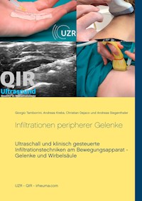
Ultrasound and clinically guided Injection techniques on the musculoskeletal system E-Book
Giorgio Tamborrini
62,99 €
Mehr erfahren.
- Herausgeber: Books on Demand
- Kategorie: Fachliteratur
- Sprache: Englisch
Targeted therapeutic injections of joints, periarticular structures, tendon sheaths or bursae are among the most important treatments for inflammatory rheumatic or inflammatory activated degenerative diseases of the musculoskeletal system. Furthermore, injections of periarticular structures are also performed in post-traumatic situations or in overuse syndromes. In this publication, we discuss indications, contraindications, the clinical and ultrasound-guided techniques preferred by the authors, and possible adverse drug reactions and side effects of intraarticular and periarticular injection.
Sie lesen das E-Book in den Legimi-Apps auf:
Seitenzahl: 85
Veröffentlichungsjahr: 2019
Ähnliche


Authors
KD Dr. med. Giorgio Tamborrini
Rheumatology FMH
Ultrasound QIR / SGUM / EFSUMB / EULAR
Interventional Pain Medicine SSIPM
Clinical Lecturer (KD) University of Zurich
Ultrasound Center Rheumatology Basel
University Hospital Basel, Switzerland
Assoz. Prof. Priv. Doz. Dr. med. univ. Christian Dejaco, Ph.D., MBA
Dienst für Rheumatologie | Servizio di reumatologia
Südtiroler Sanitätsbetrieb | Azienda Sanitaria dell'Alto Adige
Krankenhaus Bruneck | Ospedale di Brunico
Spitalstraße 11 | Via Ospedale 11
39031 Bruneck | Brunico, Italy
Dr. med. George Bruyn, MD, PhD
Consultant rheumatologist
MC Groep Hospitals
Lelystad, Netherlands
PD Dr. med. Andreas Siegenthaler
Anaesthesiology FMH
Interventional Pain Medicine SSIPM
Lindenhof Hospital, Berne, Switzerland
table of contents
Introduction
Technique
Needles
Drugs
Contraindications, complications and side effects
Musculoskeletal Ultrasound
Shoulder
7.1 Sternoclavicular joint
7.1.1 Ultrasound Sternoclavicular Joint
7.2 AC Joint (Acromioclavicular Joint)
7.2.1 Ultrasound AC-joint
7.3 Glenohumeral joint
7.3.1 Glenohumeral joint injection from anterior
7.3.2 Glenohumeral injection from posterior
7.3.3 Ultrasound Glenohumeral Joint
7.4 Bursa subdeltoidea
7.4.1 Injection from posterior
7.4.2 Lateral injection
7.4.3 Ultrasound Bursa subdeltoidea
7.5 Biceps tendon recess
7.5.1 Ultrasound biceps recess
7.6 Coracoid process
7.6.1 Ultrasound coracoid process
Elbow
8.1 Elbow joint
8.1.1 Lateral injection
8.1.2 Injection from posterior
8.1.3 Ultrasound elbow joint
8.2 Medial and lateral epicondyle
8.2.1 Injection lateral epicondyle
8.2.2 Injection medial epicondyle
8.2.3 Ultrasound medial and lateral epicondyle
8.3 Olecranon bursa
8.3.1 Ultrasound Olecranon bursa
8.4 Bicipitoradial bursa
8.4.1 Ultrasound of the bicipitoradial bursa
Hand
9.1 Wrist
9.1.1 Injection of distal radioulnar joint
9.1.2 Injection of the radiocarpal joint
9.1.3 Ulnocarpal joint injection
9.1.4 Injection of intercarpal joints and carpometacarpal joints
9.1.5 Ultrasound wrist
9.2 Metacarpophalangeal joint (MCP joint)
9.2.1 Ultrasound MCP joint
9.3 Proximal Interphalangeal Joint (PIP Joint)
9.3.1 Ultrasound PIP Joint
9.4 Distal Interphalangeal Joint (DIP Joint)
9.4.1 Ultrasound DIP Joint
9.5 Carpometacarpal joint I (CMC-I joint; thumb saddle joint)
9.5.1 Ultrasound CMC-1 joint
9.6 Extensor tendons
9.6.1 Ultrasound extensor tendons
9.7 Flexor tendons
9.7.1 Ultrasound flexor tendons
9.8 Carpal tunnel
9.8.1 Ultrasound carpal tunnel
Hip
10.1 Hip joint
10.1.1 Ultrasound hip joint
10.2.1 Ultrasound greater trochanter
10.3 Lateral femoral cutaneous nerve (LFCN)
10.3.1 Ultrasound LFCN
Knee
11.1 Knee joint
11.1.1 Ultrasound knee joint
11.2 Bursae at the knee
11.2.1 Ultrasound of the bursae at the knee
11.3 Pes anserine
11.3.1 Ultrasound Pes anserinus
Foot
12.1 Ankle joint (tibio-talar joint)
12.1.1 Ultrasound ankle joint
12.2 Subtalar joint
12.2.1 Ultrasound subtalar joint
12.3 Midfoot joints
12.4 Metatarsophalangeal Joint I
12.4.1 Ultrasound MTP Joint
12.5 Interphalangeal joints (IP joints)
12.5.1 Ultrasound IP-joints
12.6 Intermetatarsal space
12.6.1 Ultrasound intermetatarsal space
12.7 Tendon sheaths
12.7.1 Medial ankle tendon sheaths
12.7.2 Ultrasound medial tendon sheaths
12.7.3 Tarsal Tunnel
12.7.4 Ultrasound tarsal tunnel
12.7.5 Lateral tendon sheaths
12.7.6 Ultrasound of the lateral ankle tendon sheaths
12.7.7 Anterior ankle tendon sheaths
12.7.8 Ultrasound of anterior tendon sheaths
12.8 Bursa retrocalcanea
12.8.1 Ultrasound Bursa retrocalcanea
12.9 Plantar fascia
12.9.1 Plantar fascia ultrasound
Temporomandibular joint
13.1.1 Ultrasound of the temporomandibular joint
Spine and selected nerves
14.1. Cervical Facet Joints and Medial Branches
14.2. Lumbar Facet Joints and Medial Branches
14.3. Caudal epidural injection
14.4. Sacroiliac joint injection
14.5. Block of the Greater Occipital Nerve (GON)
14.6. Cervical Nerve Root Block
14.7. Stellate Ganglion Block
14.8. Block of the Ilioinguinal and Iliohypogastric nerve
14.9. Lateral cutaneus femoral nerve block (another variant)
Literature
1 Introduction
Targeted therapeutic injections of joints, periarticular structures, tendon sheaths or bursae are among the most important treatments for inflammatory rheumatic or inflammatory activated degenerative diseases of the musculoskeletal system. Furthermore, injections of periarticular structures are also performed in post-traumatic situations or in overuse syndromes (Table 1).
Indications for injections of the musculoskeletal system
Table 1
In the case of articular or periarticular inflammation, targeted local injection enables rapid anti-inflammatory action with few side effects, which often lasts for a long time in the case of arthritis, for example, through the use of crystalline steroid preparations.
If an effusion is present, it often makes sense to perform a diagnostic aspiration and/or therapeutic injection with determination of cell count, examination of the gram preparation, cell differentiation, crystal analysis and, depending on the clinic, culture and PCR examinations before injection. Synovial fluid analysis helps to differentiate between non-inflammatory and inflammatory arthropathy (Table 2).
Normal
non-inflammatory
inflammatory
septic
haemorrhagic
Colour
clear
clear
murky
murky
bloody
Viscosity
high
high
low
diverse
diverse
wbc/mm3
<200
≤2000
>2000
>50000
diverse
%PMN
<25
≤25
>25
>50
diverse
In this publication, we discuss indications, contraindications, the clinical and ultrasound-guided techniques preferred by the authors, and possible adverse drug reactions and side effects of intraarticular and periarticular injection.
2 Technique
The requirements for a correct injection technique are a clear (or suspected) diagnosis (if infection is suspected, no injection but only diagnostic aspiration ist done), an adequate information and informed patient consent (according to local national guidelines), musculoskeletal ultrasound and profound anatomical knowledge, the correct dosage of medication and, in particular, sufficient practical skills in carrying out injections. For injections in anatomically more difficult regions or for diagnostic injection of only small quantities of effusions, orientation through high-resolution musculoskeletal ultrasound (hrMSUS) is indispensable today. The hrMSUS can be used for pre-interventional exact localization of the injection site or for performing injection under direct view using various techniques.
A diagnostic injection or therapeutic injection should be painless or almost painless. The intervention takes place in a clean room without draughts, the patient should be comfortable and relaxed positioned.
In the clinically guided injection technique, we orient ourselves on the surface anatomy, especially using the osseous landmarks. The injection site is marked after orienting palpation by gentle pressure with an unsoiled ballpoint pen tip (no coloured marking) (Fig. 1).
Fig. 1: Marking of the injection site with a ballpoint pen
The injection site should not be in the area of a skin rash or a blood vessel. Shaving a hairy area is not necessary. The injection site is then disinfected according to the instructions of the product used. The skin can be superficially cryoanesthetized at the injection site before disinfection using sterile ice spray. A prior injection anaesthesia with a local anaesthetic is not necessary with the correct technique and doing a rapid injection. For special interventions, e.g. in the case of needling of a calcification of the rotator cuff of the shoulder, we recommend the prior application of a local anaesthetic into the subdeltoid bursa. In the case of several injections, e.g. at finger joints, a block anaesthesia can be evaluated. In children, the prior application of a local anaesthetic ointment or patch may be helpful.
Wearing a surgical mask is recommended, but the use of sterile gloves is not mandatory if a sterile no-touch technique e.g. according to the guidelines of the Swiss Society for Rheumatology (Tab. 3) is followed. The illustrations and pictures in this manual were made without wearing gloves.
Requirements for correct injection of the musculoskeletal system
* Informing the patient about the purpose of the injection and possible side effects
* Clean room
* Surgical mask
* Non-sterile gloves
* Use of disposable material
* Wipe disinfection with approved disinfectant, observe correct exposure time
* No-touch injection technique
Tab. 3.
When injecting a joint after clinical or prior sonographic orientation, the injection is usually made perpendicular to the skin surface. In paratendinous injections or directly (real-time) ultrasound-guided injections, a flatter insertion angle is selected depending on the structure to be injected. Ideally, with each "blind" intra-articular injection, synovial fluid is aspirated before an injection is performed, which proves the safe intraarticular position of the needle (not necessary with the direct ultrasound-guided injection technique). As mentioned above, aspiration of synovial fluid enables diagnostic analysis and leads to therapeutic relief in the case of large quantities of an effusion. The injection of a drug should be done without resistance and painless. After the injection, a short compression of the injection site with a sterile swab and a sterile adhesive plaster should be applied.
By following this procedure, injection will be fast, safe and efficient. In case of insufficient success, a second injection can be carried out after 2-4 weeks.
3 Needles
The suitable disposable injection needle (Fig. 2), the injection volume and the applied dose of medication depend on the size and position of the joint and are mentioned individually for each joint (Table 4


